血管扩张Angiectasis · 阴蒂腺 · 雌性生殖系统 · NTP ntp-0557
诊断要点:阴蒂腺——血管扩张:只要存在即应作出诊断并分级。
病变描述
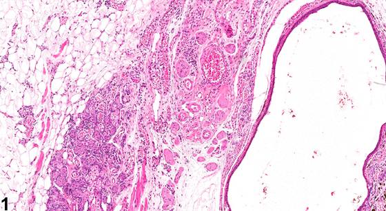
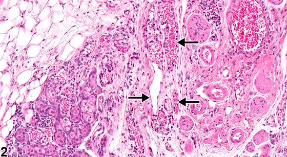
在血管扩张(angiectasis)中,原有的阴蒂腺血管扩张并充满血细胞,伴有阴蒂腺实质的受压(Figure 1、Figure 2)。应尝试区分血管扩张与血管瘤(hemangioma),尽管这种区分并不总是显而易见。血管瘤往往为边界清楚、无包膜的肿块,由紧密排列的扩张血管腔隙构成。每个血管腔隙均由单层形态正常的内皮细胞围成并衬覆,这些内皮细胞排列于胶原性间隔上,间隔通常较薄,但部分具有宽阔的胶原性间质。血管扩张通常不表现为边界清楚的肿块:扩张的血管通道常不规则地穿行于组织中。血管扩张和血管瘤均可发生邻近实质的受压。
诊断与分级建议(NTP 共识)
阴蒂腺——血管扩张:只要存在即应作出诊断并分级。
图谱
英文原文
展开 NTP 官方英文描述与建议
Comment:
In angiectasis, preexisting clitoral gland blood vessels become dilated and filled with blood cells, accompanied by compression of the clitoral gland parenchyma ( Figure 1 and Figure 2 ). A distinction between angiectasis and hemangioma should be attempted, although the distinction is not always obvious. Hemangiomas tend to be well-circumscribed, unencapsulated masses composed of tightly packed, dilated vascular spaces. Each vascular space is enclosed and lined by a single layer of normal-appearing endothelial cells aligned on collagenous septa, which are usually thin, although some have broad collagenous stroma. Angiectasis does not usually present as a well-circumscribed mass: the dilated vascular channels often course irregularly through the tissue. Compression of adjacent parenchyma may occur in both angiectasis and hemangioma.
Recommendation:
Clitoral gland - Angiectasis should be diagnosed and graded whenever present.
来源与引用
原始条目:https://ntp.niehs.nih.gov/atlas/nnl/reproductive-system-female/clitoral-gland/Angiectasis
图谱总目录:NTP Nonneoplastic Lesion Atlas。 内容属美国联邦政府作品(公有领域),可引用;引用时请标注来源。
建议引用格式:NTP Nonneoplastic Lesion Atlas. Angiectasis (clitoral-gland). LabSmart 中文版:
https://www.labsmart.cn/ntp/clitoral-gland-angiectasis.html
中文图谱总页(可展开对照全部 595 条):https://www.labsmart.cn/ntp-atlas.html#clitoral-gland-angiectasis