萎缩Atrophy · 腺胃 · 消化系统 · NTP ntp-0283
诊断要点:萎缩应当诊断并分级。严重程度分级取决于病变范围及上皮细胞丢失/体积减小的程度。
病变描述
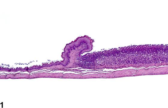
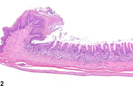
腺胃病变在NTP小鼠中罕见。以黏膜上皮细胞体积减小和数量减少为特征的局灶性(focal)黏膜萎缩可能由溃疡、炎症、矿化或梗死引起。广泛性黏膜萎缩可如Figure 2、Figure 4所示由化学物质诱导。弥漫性(diffuse)萎缩也可为年龄相关变化,表现为黏膜高度弥漫性降低和黏膜纤维结缔组织增多。在老龄大鼠中,胃底腺可呈弥漫性萎缩,壁细胞和主细胞减少,并含有囊肿。腺体被纤维结缔组织取代,固有层和腺上皮内伴有局灶性淋巴细胞浸润,沿界嵴伴有混合性炎症细胞浸润。
诊断与分级建议(NTP 共识)
萎缩应当诊断并分级。严重程度分级取决于病变范围及上皮细胞丢失/体积减小的程度。
图谱
英文原文
展开 NTP 官方英文描述与建议
Comment:
Lesions of the glandular stomach are rare in NTP mice. Focal mucosal atrophy characterized by decreased size and number of mucosal epithelial cells may result from ulceration, inflammation, mineralization, or infarction. Generalized mucosal atrophy can be chemically induced as in Figure 2 and Figure 4 . Diffuse atrophy can also be an age-related change with a diffuse decrease of mucosal height and increased mucosal fibrous connective tissue. In the aged rat, fundic glands may be diffusely atrophic, with fewer parietal and chief cells, and contain cysts. Glands are replaced by fibrous connective tissue with focal lymphocytic infiltrates in the lamina propria and the glandular epithelium and a mixed inflammatory infiltrate along the limiting ridge.
Recommendation:
Atrophy should be diagnosed and graded. Severity grade depends on the extent of the lesion and degree of loss/decreased size of epithelial cells.
来源与引用
原始条目:https://ntp.niehs.nih.gov/atlas/nnl/alimentary-system/stomach-glandular-stomach/Atrophy
图谱总目录:NTP Nonneoplastic Lesion Atlas。 内容属美国联邦政府作品(公有领域),可引用;引用时请标注来源。
建议引用格式:NTP Nonneoplastic Lesion Atlas. Atrophy (stomach-glandular-stomach). LabSmart 中文版:
https://www.labsmart.cn/ntp/stomach-glandular-stomach-atrophy.html
中文图谱总页(可展开对照全部 595 条):https://www.labsmart.cn/ntp-atlas.html#stomach-glandular-stomach-atrophy