细胞浸润Infiltration Cellular · 腺胃 · 消化系统 · NTP ntp-0280
诊断要点:当炎症细胞存在而无其他炎症特征(例如水肿、出血、变性、坏死)时,应使用“infiltrate, cellular”一词。
病变描述
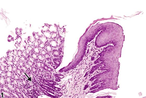
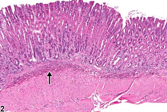
“细胞浸润(cellular infiltrate)”一词常用于描述炎症细胞的存在,而无其他炎症过程的证据(例如水肿、坏死或细胞变性、血管损伤的证据)。浸润通常由淋巴细胞、肥大细胞或嗜酸性粒细胞组成。腺胃中的淋巴细胞浸润可为偶然发现;它们通常出现在黏膜深层和黏膜下层。在图1(箭头)中,淋巴细胞浸润存在于腺胃深部黏膜和前胃固有层。嗜酸性粒细胞(图2和图3,箭头)在过敏性疾病和寄生虫病中存在的嗜酸性粒细胞趋化因子作用下从血流被募集到大多数器官,并提示超敏反应。胃黏膜下层偶可见肥大细胞的局灶性(focal)聚集。肥大细胞已被证明在急性过敏反应、迟发相反应和慢性过敏性炎症中起重要作用。胃肠道黏膜下层肥大细胞的局灶性聚集在NTP研究中通常被视为偶然性病变。此类聚集可能与肥大细胞瘤混淆。肥大细胞的局灶性聚集往往比肥大细胞瘤更小且边界更不清楚,并且浸润于结构之间而非压迫或取代邻近结构。
诊断与分级建议(NTP 共识)
当炎症细胞存在而无其他炎症特征(例如水肿、出血、变性、坏死)时,应使用“infiltrate, cellular”一词。一般而言,炎症细胞浸润往往相当小,可能不足以作出诊断。当作出诊断时,应根据细胞数量和受累组织总面积对浸润进行分级。应使用修饰词指明具体的细胞类型(例如淋巴细胞、嗜酸性粒细胞、肥大细胞)。
图谱
英文原文
展开 NTP 官方英文描述与建议
Comment:
The term "cellular infiltrate" is often used to describe the presence of inflammatory cells without other evidence of an inflammatory process (e.g., edema, necrosis or degeneration of cells, evidence of vascular injury). Infiltrates usually consist of lymphocytes, mast cells, or eosinophils. Lymphocytic infiltrates in the glandular stomach can be an incidental finding; they typically appear in the deep aspect of the mucosa and in the submucosa. In Figure 1 (arrow), lymphocytic infiltrates are present in the deep mucosa of the glandular stomach and in the propria of the forestomach. Eosinophils ( Figure 2 and Figure 3 , arrows) are recruited from the blood stream into most organs in response to eosinophil chemoattractants present in allergic and parasitic diseases and indicate a hypersensitivity reaction. Focal collections of mast cells can occasionally be found in the submucosa of the stomach. Mast cells have been shown to contribute importantly to acute allergic reactions, late-phase reactions, and chronic allergic inflammation. Focal accumulations of mast cells in the submucosa of the gastrointestinal tract are usually considered an incidental lesion in NTP studies. Such accumulations may be confused with mast cell tumor. Focal accumulations of mast cells tend to be smaller and less well circumscribed than mast cell tumors and infiltrate between structures rather than compressing or effacing adjacent structures.
Recommendation:
The term "infiltrate, cellular" should be used when inflammatory cells are present with no other features of inflammation (e.g., edema, hemorrhage, degeneration, necrosis). Generally, infiltrates of inflammatory cells are often quite small and may not warrant a diagnosis. When diagnosed, infiltrates should be graded based on the number of cells and the total tissue area affected. A modifier should be used to indicate the specific type of cell (e.g., lymphocyte, eosinophil, mast cell).
来源与引用
原始条目:https://ntp.niehs.nih.gov/atlas/nnl/alimentary-system/stomach-glandular-stomach/InfiltrationCellular
图谱总目录:NTP Nonneoplastic Lesion Atlas。 内容属美国联邦政府作品(公有领域),可引用;引用时请标注来源。
建议引用格式:NTP Nonneoplastic Lesion Atlas. Infiltration Cellular (stomach-glandular-stomach). LabSmart 中文版:
https://www.labsmart.cn/ntp/stomach-glandular-stomach-infiltration-cellular.html
中文图谱总页(可展开对照全部 595 条):https://www.labsmart.cn/ntp-atlas.html#stomach-glandular-stomach-infiltration-cellular