矿化Mineralization · 腺胃 · 消化系统 · NTP ntp-0276
诊断要点:应当诊断矿化并根据矿化范围进行分级。继发于矿化的相关病变,如炎症或胃上皮细胞变性,不应单独诊断,而应在叙述中描述。
病变描述
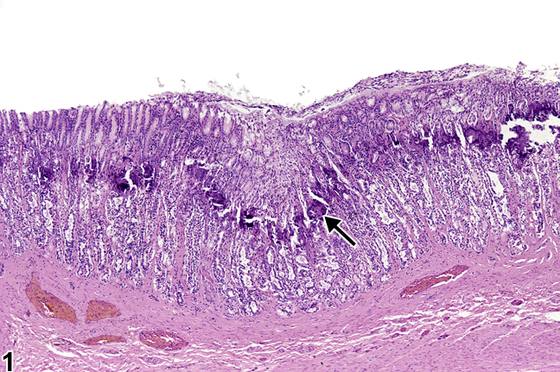
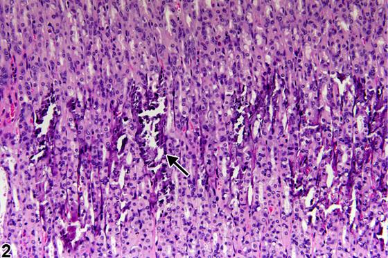
以局灶性聚集体或致密蓝染矿物质凝结物为特征的矿化,在老年大鼠胃的H&E染色切片中相当常见,此类矿化与胃腺的囊性扩张相关;小鼠和仓鼠偶尔也可见类似变化。当啮齿类动物存在矿物质代谢紊乱时,胃矿化可能变得显著(显著(marked)),尤其是在慢性进行性肾病和尿毒症时,这是由于钙磷代谢改变和甲状旁腺功能亢进所致。这些变化的特征为整个胃底、肌层和间质的血管发生矿化,呈与黏膜富含壁细胞区域平行的带状分布(Figure 1、Figure 2,箭头所示),但不累及贲门、胃窦或幽门。矿物质沉积物在环绕上皮和血管的基底膜周围形成。固有层因水肿而扩张,并出现间质纤维增生。胃腺扩张,伴壁细胞肿胀和变性以及主细胞萎缩。发生腺上皮糜烂伴出血。胃矿化常伴有黏液细胞增生。
诊断与分级建议(NTP 共识)
应当诊断矿化并根据矿化范围进行分级。继发于矿化的相关病变,如炎症或胃上皮细胞变性,不应单独诊断,而应在叙述中描述。
图谱
英文原文
展开 NTP 官方英文描述与建议
Comment:
Mineralization characterized by focal aggregates or concretions of densely blue-staining mineral is fairly common in the H&E-stained sections from the stomachs of aged rats, where they are associated with cystic dilation of the gastric glands; mice and hamsters occasionally show similar changes. Gastric mineralization may become marked in rodents when there is disturbance in mineral metabolism, particularly with chronic progressive renal disease and uremia due to altered calcium-phosphorus metabolism and hyperparathyroidism. These changes are characterized by mineralization of the vessels throughout the gastric fundus, muscular tunics, and interstitium in a band paralleling the parietal-cell-rich region of the mucosa ( Figure 1 and Figure 2 , arrows) but not the cardia, antrum, or pylorus. Mineral deposits develop around basement membranes surrounding epithelium and blood vessels. The lamina propria becomes expanded by edema, and fibroplasia of the interstitium develops. The gastric glands become dilated with swelling and degeneration of parietal cells and atrophy of chief cells. Erosion of the glandular epithelium with hemorrhage occurs. Gastric mineralization is frequently accompanied by mucous cell hyperplasia.
Recommendation:
Mineralization should be diagnosed and graded depending on the extent of mineralization. Associated lesions, such as inflammation or degeneration of gastric epithelial cells, that are secondary to the mineralization should not be diagnosed separately but should be described in the narrative.
来源与引用
原始条目:https://ntp.niehs.nih.gov/atlas/nnl/alimentary-system/stomach-glandular-stomach/Mineralization
图谱总目录:NTP Nonneoplastic Lesion Atlas。 内容属美国联邦政府作品(公有领域),可引用;引用时请标注来源。
建议引用格式:NTP Nonneoplastic Lesion Atlas. Mineralization (stomach-glandular-stomach). LabSmart 中文版:
https://www.labsmart.cn/ntp/stomach-glandular-stomach-mineralization.html
中文图谱总页(可展开对照全部 595 条):https://www.labsmart.cn/ntp-atlas.html#stomach-glandular-stomach-mineralization