切牙变性Incisor Degeneration · 牙齿 · 消化系统 · NTP ntp-0248
诊断要点:牙齿,门齿 - 退行性变:只要存在即应诊断并分级。如果牙本质层外观正常而仅成牙本质细胞发生退行性变或坏死,则应诊断为“牙齿,成牙本质细胞 - 退行性变”或“牙齿,成牙本质细胞 - 坏死”。
病变描述
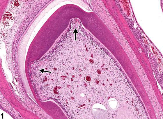
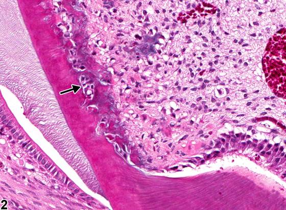
门齿的退行性改变在NTP研究中不常见。门齿退行性变包括一组病变,这些病变可能不会同时出现。早期病变包括牙髓内局灶性(focal)间充质细胞空泡化和骨牙本质形成。更严重的病变包括局灶性或多灶性(multifocal)成牙本质细胞退行性变伴牙本质龛形成(Figure 1、Figure 2)、牙本质基质排列异常以及基质染色改变(Figure 3、Figure 4)。严重病变中可见牙本质层断裂。门齿退行性变可能由化学物质诱导,并且至少在大鼠的一个案例中,有研究提示其继发于选择性前成牙本质细胞群损伤。
诊断与分级建议(NTP 共识)
牙齿,门齿 - 退行性变:只要存在即应诊断并分级。如果牙本质层外观正常而仅成牙本质细胞发生退行性变或坏死,则应诊断为“牙齿,成牙本质细胞 - 退行性变”或“牙齿,成牙本质细胞 - 坏死”。
图谱
英文原文
展开 NTP 官方英文描述与建议
Comment:
Degenerative changes of the incisors are uncommon in NTP studies. Incisor degeneration comprises a set of lesions that may not all be present concurrently. Early lesions include focal mesenchymal cell vacuolation and osteodentin formation within the pulp. More severe lesions include focal or multifocal odontoblast degeneration with dentin niche formation ( Figure 1 and Figure 2 ), abnormal organization of the dentin matrix, and altered matrix staining ( Figure 3 and Figure 4 ). Breaks in the dentin layer may be seen with severe lesions. Incisor degeneration may be chemically induced and, at least in one case in rats, has been suggested to be secondary to injury to a selective population of preodontoblasts.
Recommendation:
Tooth, Incisor - Degeneration should be diagnosed and graded whenever present. If the dentin layer appears normal and the odontoblasts only are undergoing degeneration or necrosis, then "tooth, odontoblast - degeneration" or "tooth, odontoblast - necrosis" should be diagnosed.
来源与引用
原始条目:https://ntp.niehs.nih.gov/atlas/nnl/alimentary-system/tooth/Incisor-Degeneration
图谱总目录:NTP Nonneoplastic Lesion Atlas。 内容属美国联邦政府作品(公有领域),可引用;引用时请标注来源。
建议引用格式:NTP Nonneoplastic Lesion Atlas. Incisor Degeneration (tooth). LabSmart 中文版:
https://www.labsmart.cn/ntp/tooth-incisor-degeneration.html
中文图谱总页(可展开对照全部 595 条):https://www.labsmart.cn/ntp-atlas.html#tooth-incisor-degeneration