淋巴细胞增生Hyperplasia Lymphocyte · 淋巴结 · 免疫系统 · NTP ntp-0006
诊断要点:淋巴结淋巴细胞增生无论何时出现,均应作出诊断、标明部位(如滤泡、副皮质区)并分级。
病变描述
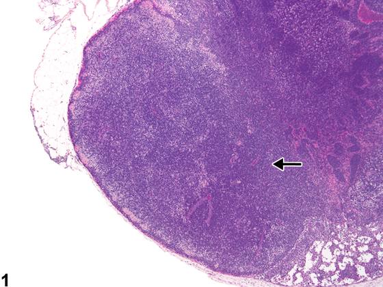
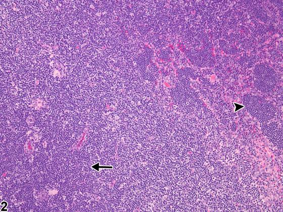
在啮齿类动物中,淋巴细胞增生(lymphocyte hyperplasia)可能累及淋巴结的多个解剖区域,包括富含B细胞的滤泡(滤泡性)、富含T细胞的副皮质区(副皮质性)、髓索,或三者全部受累。滤泡性增生(follicular hyperplasia)表现为初级滤泡数量和体积增加,有时还包括刺激形成次级滤泡(生发中心)。受刺激的生发中心含有大的淋巴母细胞(中心母细胞),并伴有淋巴细胞凋亡增加及可染体巨噬细胞。副皮质区淋巴细胞增生(paracortical lymphocyte hyperplasia)表现为细胞密度增加,偶伴面积增大(Figure 1、Figure 2,箭头)。淋巴细胞增生可伴有血管内淋巴细胞数量增加(淋巴细胞运输)(Figure 2,箭头头)。淋巴细胞增生在正常啮齿类动物中可呈现不同程度,取决于部位(如肠系膜或下颌淋巴结)、年龄、动物健康状态及切面。该病变通常被认为是反应性或免疫反应,而非肿瘤前病变。淋巴细胞增生应与淋巴瘤相鉴别。增生时,淋巴细胞成熟、小、形态正常,并存在于各自相应的区域;而淋巴瘤通常以肿瘤性淋巴细胞呈单一形态群体、淋巴结结构破坏、淋巴结体积增大及侵犯邻近组织为特征。
诊断与分级建议(NTP 共识)
淋巴结淋巴细胞增生无论何时出现,均应作出诊断、标明部位(如滤泡、副皮质区)并分级。由于淋巴结存在变异性,淋巴细胞增生仅应在仔细评估并与同期对照比较后作出诊断。诊断该病变时必须考虑切片的充分性(即浅表或切向切片)。对于某些淋巴结(如腘淋巴结),可能难以获得充分的切片。
图谱
英文原文
展开 NTP 官方英文描述与建议
Comment:
In rodents, lymphocyte hyperplasia may involve various anatomical compartments of the lymph nodes, including the B-cell–rich follicles (follicular), the T-cell-rich paracortex (paracortical), the medullary cords, or all three. Follicular hyperplasia is identified by an increase in number and size of primary follicles and sometimes includes stimulation that results in secondary follicles (germinal centers). Stimulated germinal centers contain large lymphoblasts (centroblasts) and increased lymphocyte apoptosis with tingible-body macrophages. Paracortical lymphocyte hyperplasia is identified by increased cell density, occasionally accompanied by increased area ( Figure 1 and Figure 2 , arrows). Lymphocyte hyperplasia may be accompanied by increased numbers of lymphocytes within vessels (trafficking) of lymphocytes ( Figure 2 , arrowhead). Lymphocyte hyperplasia can be evident to varying degrees in normal rodents, depending on location (e.g., mesenteric or mandibular lymph nodes), age, animal health status, and the plane of section. This lesion is generally considered to be a reactive or immune response and not a preneoplastic lesion. Lymphocyte hyperplasia should be distinguished from lymphoma. With hyperplasia, lymphocytes are mature, small, and normal in appearance and present within their respective compartments, whereas lymphoma typically features a monomorphic population of neoplastic lymphocytes, effacement of the nodal architecture, increased lymph node size, and invasion of adjacent tissue.
Recommendation:
Whenever present, lymphocyte hyperplasia of lymph nodes should be diagnosed, localized (e.g., follicle, paracortex), and graded. Due to lymph node variability, lymphocyte hyperplasia should be diagnosed only after careful evaluation and comparison with concurrent controls. Adequacy of the section (i.e., superficial or tangential) must be taken into consideration in the diagnosis of this lesion. For some lymph nodes (e.g., popliteal), an adequate section may be difficult to obtain.
来源与引用
原始条目:https://ntp.niehs.nih.gov/atlas/nnl/immune-system/lymph-node/HyperplasiaLymphocyte
图谱总目录:NTP Nonneoplastic Lesion Atlas。 内容属美国联邦政府作品(公有领域),可引用;引用时请标注来源。
建议引用格式:NTP Nonneoplastic Lesion Atlas. Hyperplasia Lymphocyte (lymph-node). LabSmart 中文版:
https://www.labsmart.cn/ntp/lymph-node-hyperplasia-lymphocyte.html
中文图谱总页(可展开对照全部 595 条):https://www.labsmart.cn/ntp-atlas.html#lymph-node-hyperplasia-lymphocyte