浆细胞增生Hyperplasia Plasma Cell · 淋巴结 · 免疫系统 · NTP ntp-0003
诊断要点:淋巴结中的浆细胞增生应当被诊断并给出严重程度分级。然而,如果该病变伴随淋巴细胞增生和/或肿瘤性病变,则不应单独诊断,而应在叙述性描述中加以描述。
病变描述
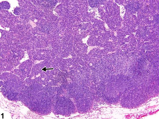
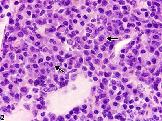
浆细胞增生(plasma cell hyperplasia)是啮齿类动物淋巴结中的常见病变,尤其多见于下颌淋巴结(mandibular lymph nodes)(Figure 1、Figure 2),最常见的原因是接触饮食性抗原后的反应。如果不是背景病变,则浆细胞增生可能与受试物相关,或与其他器官的炎性、感染性或肿瘤性病变相关。浆细胞及其前体细胞正常情况下存在于髓索(medullary cords)中;因此,髓索是浆细胞增生的主要部位(Figure 1)。浆细胞通常因需要产生抗体的抗原刺激而增多,因此通常B细胞(淋巴样)增生伴有浆细胞增生。髓系增生(myeloid hyperplasia)也可能存在。成熟浆细胞胞质呈嗜碱性至嗜酸性,细胞核偏位,可见高尔基体透明区(Golgi clear zone),染色质呈车轮状(cartwheel chromatin pattern)(Figure 2,箭头)。根据抗原刺激的程度和慢性程度,可能出现一些含有拉塞尔小体(Russell bodies)的莫特细胞(Mott cells)。在显著(marked)浆细胞增生时,淋巴结可显著增大,主要由浆细胞构成,并可能表现为正常淋巴结结构的部分消失。显著浆细胞增生可通过后者(浆细胞肿瘤)中存在浆细胞前体细胞、非典型浆细胞、被膜浸润和转移而与浆细胞肿瘤相鉴别。
诊断与分级建议(NTP 共识)
淋巴结中的浆细胞增生应当被诊断并给出严重程度分级。然而,如果该病变伴随淋巴细胞增生和/或肿瘤性病变,则不应单独诊断,而应在叙述性描述中加以描述。
图谱
英文原文
展开 NTP 官方英文描述与建议
Comment:
Plasma cell hyperplasia is a common finding in the lymph nodes of rodents, particularly in the mandibular lymph nodes ( Figure 1 and Figure 2 ), most often in response to exposure to dietary antigens. If not a background lesion, then plasma cell hyperplasia may be treatment related or associated with inflammatory, infectious, or neoplastic lesions in other organs. Plasma cells and their precursors are normally present in the medullary cords; therefore, these cords are the primary site of plasma cell hyperplasia ( Figure 1 ). Plasma cells usually increase in response to antigenic stimulation requiring antibody production, so typically B-cell (lymphoid) hyperplasia is accompanied by plasma cell hyperplasia. Myeloid hyperplasia may also be present. Mature plasma cells have basophilic to eosinophilic cytoplasm, eccentrically located nuclei, a Golgi clear zone, and a cartwheel chromatin pattern ( Figure 2 , arrows). Depending on the degree and chronicity of antigenic stimulation, some Mott cells with Russell bodies may be present. With marked plasma cell hyperplasia, the lymph node may be greatly enlarged and composed mostly of plasma cells and may exhibit partial effacement of normal nodal architecture. Marked plasma cell hyperplasia may be differentiated from plasma cell neoplasia by the presence of plasma cell precursors, atypical plasma cells, capsular infiltration, and metastases in the latter.
Recommendation:
Plasma cell hyperplasia in lymph nodes should be diagnosed and given a severity grade. However, if this lesion accompanies lymphocyte hyperplasia and/or neoplasia, it should not be diagnosed separately but should be described in the narrative.
来源与引用
原始条目:https://ntp.niehs.nih.gov/atlas/nnl/immune-system/lymph-node/HyperplasiaPlasmaCell
图谱总目录:NTP Nonneoplastic Lesion Atlas。 内容属美国联邦政府作品(公有领域),可引用;引用时请标注来源。
建议引用格式:NTP Nonneoplastic Lesion Atlas. Hyperplasia Plasma Cell (lymph-node). LabSmart 中文版:
https://www.labsmart.cn/ntp/lymph-node-hyperplasia-plasma-cell.html
中文图谱总页(可展开对照全部 595 条):https://www.labsmart.cn/ntp-atlas.html#lymph-node-hyperplasia-plasma-cell