上皮增生Epithelium Hyperplasia · 卵巢 · 雌性生殖系统 · NTP ntp-0505
诊断要点:卵巢,上皮——增生应始终予以诊断并给出严重程度分级。
病变描述
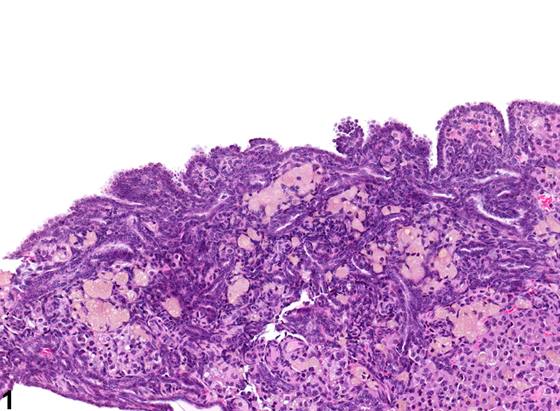
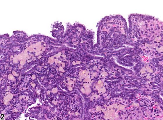
卵巢表面上皮增生表现为上皮表面的小息肉样突起,以及由胞质嗜碱性(basophilic)的立方上皮细胞索向下生长(Figure 1、Figure 2)。上皮细胞增生被认为起源于覆盖卵巢表面的间皮(mesothelium)。过去,这被称为生发上皮(germinal epithelium),但“生发上皮”一词具有误导性,因为覆盖卵巢表面的上皮是腹膜(peritoneum)的延续。上皮细胞增生必须与腺瘤(adenoma)、囊腺瘤(cystadenoma)和肾小管间质肿瘤(tubulostromal neoplasms)相鉴别,也必须与管状(Sertoli细胞样)增生(tubular (Sertoliform) hyperplasia)相鉴别,后者由柱状细胞衬覆的管状结构组成,细胞质边界不清,细胞核位于基底部。
诊断与分级建议(NTP 共识)
卵巢,上皮——增生应始终予以诊断并给出严重程度分级。
图谱
英文原文
展开 NTP 官方英文描述与建议
Comment:
Proliferation of the epithelium lining the surface of the ovary appears as small polypoid projections from the epithelial surface and as downgrowths of cords of cuboidal epithelium cells with basophilic cytoplasm ( Figure 1 and Figure 2 ). Epithelial cell hyperplasia is thought to be derived from the mesothelium lining the ovarian surface. In the past, this has been referred to as germinal epithelium but the term "germinal epithelium" is misleading, because the epithelium covering the surface of the ovary is a continuation of the peritoneum. Epithelial cell hyperplasia must be distinguished from adenoma, cystadenoma, and tubulostromal neoplasms and from tubular (Sertoliform) hyperplasia, which consists of tubules lined by columnar cells with indistinct cytoplasmic borders and basal nuclei.
Recommendation:
Ovary, Epithelium - Hyperplasia should always be diagnosed and given a severity grade.
来源与引用
原始条目:https://ntp.niehs.nih.gov/atlas/nnl/reproductive-system-female/ovary/Epithelium-Hyperplasia
图谱总目录:NTP Nonneoplastic Lesion Atlas。 内容属美国联邦政府作品(公有领域),可引用;引用时请标注来源。
建议引用格式:NTP Nonneoplastic Lesion Atlas. Epithelium Hyperplasia (ovary). LabSmart 中文版:
https://www.labsmart.cn/ntp/ovary-epithelium-hyperplasia.html
中文图谱总页(可展开对照全部 595 条):https://www.labsmart.cn/ntp-atlas.html#ovary-epithelium-hyperplasia