增生Hyperplasia · 垂体 · 内分泌系统 · NTP ntp-0049
诊断要点:局灶性(focal) hyperplasia一旦存在即应作出诊断并给予严重程度分级,且应在诊断中指明所累及的垂体部位(例如:Pituitary Gland, Pars distalis - Hyperplasia)。
病变描述
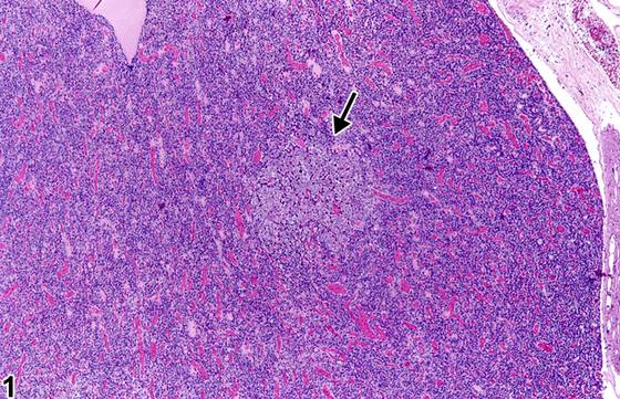
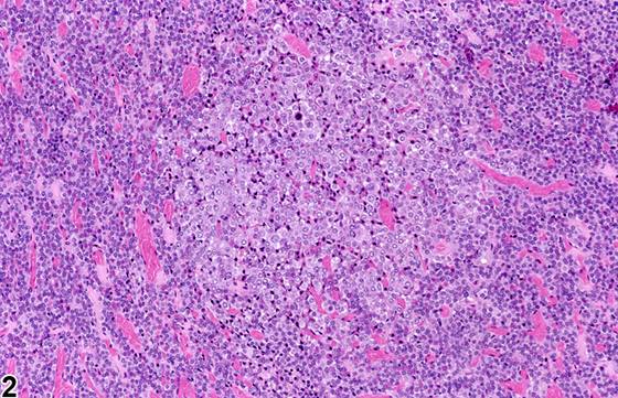
局灶性(focal) hyperplasia(局灶性增生)是一种常见的自发性及诱发性改变,在大鼠中比小鼠中更常见。其显微镜下表现多样,但主要由单一细胞类型构成,与邻近实质相融合而无压迫(Figure 1、Figure 2、Figure 3、Figure 4)。增生通常是细胞数量增加与细胞体积增大的组合,并随动物年龄增长而增加。有时可出现多个增生灶(Figure 4)。血管扩张(angiectasis)可发生于增生灶内(Figure 5),有时可引起邻近实质的轻度压迫。垂体中间部(pars intermedia)的局灶性 hyperplasia(Figure 6、Figure 7)较远侧部(pars distalis)增生少见。中间部的增生细胞与正常中间部细胞相似,由于可能没有邻近实质的压迫,局灶性增殖性病变的识别可能依赖于生长模式的改变或中间部的不对称增大。弥漫性(diffuse) hyperplasia(弥漫性增生)也可发生,必须与中间部的切向切片相鉴别。弥漫性增生通常为特定细胞类型,可通过免疫组织化学(immunohistochemistry)确认,通常代表一种生理性反应。增生与垂体肿瘤(pituitary neoplasia)之间存在形态学连续谱,邻近实质的压迫是肿瘤的主要诊断特征。针对垂体激素的免疫组织化学可用于确定细胞类型。
诊断与分级建议(NTP 共识)
局灶性(focal) hyperplasia一旦存在即应作出诊断并给予严重程度分级,且应在诊断中指明所累及的垂体部位(例如:Pituitary Gland, Pars distalis - Hyperplasia)。增生的任何显著特征均可在病理学描述(pathology narrative)中描述。增生性病变内的血管扩张不应单独诊断。
图谱
英文原文
展开 NTP 官方英文描述与建议
Comment:
Focal hyperplasia is a frequent spontaneous as well as induced change and is seen more commonly in rats than in mice. The microscopic appearance is variable but consists primarily of a single cell type that blends into the adjacent parenchyma without compression ( Figure 1 , Figure 2 , Figure 3 , and Figure 4 ). Hyperplasia is typically a combination of increased cell number and increased cell size and increases with animal age. On occasion there may be multiple foci of hyperplasia ( Figure 4 ). Angiectasis may occur within a focus of hyperplasia ( Figure 5 ) and may sometimes cause slight compression of adjacent parenchyma. Focal hyperplasia of the pars intermedia ( Figure 6 and Figure 7 ) is less common than hyperplasia of the pars distalis. The hyperplastic cells in the pars intermedia are similar to normal pars intermedia cells, and identification of a focal proliferative lesion may rely on alteration of growth pattern or asymmetrical enlargement of the pars intermedia since there may be no compression of the adjacent parenchyma. Diffuse hyperplasia may also occur and must be distinguished from a tangential section of the pars intermedia. Diffuse hyperplasia is usually of a specific cell type that can be confirmed by immunohistochemistry and typically represents a physiologic response. There is a morphologic continuum between hyperplasia and pituitary neoplasia, with compression of the adjacent parenchyma being a primary diagnostic feature of neoplasia. Immunohistochemistry for pituitary hormones can be used to determine cell type.
Recommendation:
Focal hyperplasia should be diagnosed and given a severity grade whenever present, and the part of the pituitary involved should be specified in the diagnosis (e.g., Pituitary Gland, Pars distalis - Hyperplasia). Any remarkable features of hyperplasia may be described in the pathology narrative. Angiectasis within the hyperplastic lesion should not be diagnosed separately.
来源与引用
原始条目:https://ntp.niehs.nih.gov/atlas/nnl/endocrine-system/pituitary-gland/Hyperplasia
图谱总目录:NTP Nonneoplastic Lesion Atlas。 内容属美国联邦政府作品(公有领域),可引用;引用时请标注来源。
建议引用格式:NTP Nonneoplastic Lesion Atlas. Hyperplasia (pituitary-gland). LabSmart 中文版:
https://www.labsmart.cn/ntp/pituitary-gland-hyperplasia.html
中文图谱总页(可展开对照全部 595 条):https://www.labsmart.cn/ntp-atlas.html#pituitary-gland-hyperplasia