萎缩Atrophy · 精囊 · 雄性生殖系统 · NTP ntp-0485
诊断要点:精囊萎缩应当诊断并分级。当双侧精囊均存在时,诊断应明确为双侧性,严重程度基于受累更严重的精囊。
病变描述
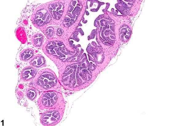
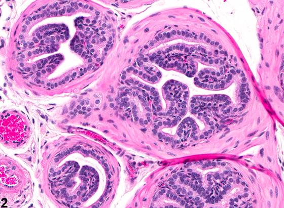
精囊萎缩(seminal vesicle atrophy)的特征是精囊腺体成分及衬覆上皮细胞的大小和数量减少。这与分泌耗竭及收缩的纤维肌性间质更加显著有关。萎缩可伴有腺体内小乳头状皱褶形成(Figure 1、Figure 2)。或者,可出现黏膜塌陷,形成由立方至低立方上皮衬覆的腺样结构(Figure 3、Figure 4)。当雄激素耗竭对包括精囊在内的附属性器官产生深刻萎缩效应时,精囊萎缩可作为年龄相关变化发生。雌激素减少腺上皮体积并增加精囊纤维肌性间质的显著性。抗雄激素物质处理也可导致精囊萎缩。在大鼠中给予多柔比星(doxorubicin)可诱导包括精囊在内的数个器官发生萎缩性改变。锌指转录因子Egr家族(如Egr4和Egr1)的缺陷,如Egr4-Egr1双突变小鼠,可导致精囊、前列腺、附睾和睾丸萎缩。
诊断与分级建议(NTP 共识)
精囊萎缩应当诊断并分级。当双侧精囊均存在时,诊断应明确为双侧性,严重程度基于受累更严重的精囊。病理学叙述中的评论应说明精囊萎缩是否因处理而加重,并应识别其他雄性生殖组织中的相关变化。
图谱
英文原文
展开 NTP 官方英文描述与建议
Comment:
Seminal vesicle atrophy is characterized by a reduction in the size and number of glandular components and lining epithelial cells of the seminal vesicle. This is associated with secretory depletion and an increased prominence of the contracted fibromuscular stroma. Atrophy may be accompanied by the formation of small papillary folds in the glands ( Figure 1 and Figure 2 ). Alternatively, there may be collapse of the mucosa, with formation of glandular-appearing structures lined by cuboidal to low cuboidal epithelium ( Figure 3 and Figure 4 ). Seminal vesicle atrophy may occur as an age-related change when the depletion of androgenic hormone has a profound atrophic effect on accessory sex organs, including the seminal vesicle. Estrogens decrease the volume of the glandular epithelium and increase the prominence of the fibromuscular stroma in the seminal vesicle. Treatment with anti-androgenic substances also leads to the atrophy of the seminal vesicle. Administration of doxorubicin in rats induces atrophic changes in several organs, including the seminal vesicle. Deficiency of the Egr family of zinc finger transcription factors like Egr4 and Egr1 as in Egr4-Egr1 double-mutant mice leads to atrophy of seminal vesicle, prostate, epididymis, and testis.
Recommendation:
Seminal vesicle atrophy should be diagnosed and graded. When present In both seminal vesicles, the diagnosis should be clarified as bilateral and severity based on the more severely affected seminal vesicle. Comments in the pathology narrative should address whether seminal vesicle atrophy is exacerbated by treatment and should identify related changes in other male reproductive tissues.
来源与引用
原始条目:https://ntp.niehs.nih.gov/atlas/nnl/reproductive-system-male/seminal-vesicle/Atrophy
图谱总目录:NTP Nonneoplastic Lesion Atlas。 内容属美国联邦政府作品(公有领域),可引用;引用时请标注来源。
建议引用格式:NTP Nonneoplastic Lesion Atlas. Atrophy (seminal-vesicle). LabSmart 中文版:
https://www.labsmart.cn/ntp/seminal-vesicle-atrophy.html
中文图谱总页(可展开对照全部 595 条):https://www.labsmart.cn/ntp-atlas.html#seminal-vesicle-atrophy