牙周袋Periodontal Pocket · 牙齿 · 消化系统 · NTP ntp-0255
诊断要点:牙周袋应当诊断,但不进行分级。牙周袋内的异物(如毛干、垫料、饲料)不应单独诊断,而应在病理描述中加以描述。
病变描述
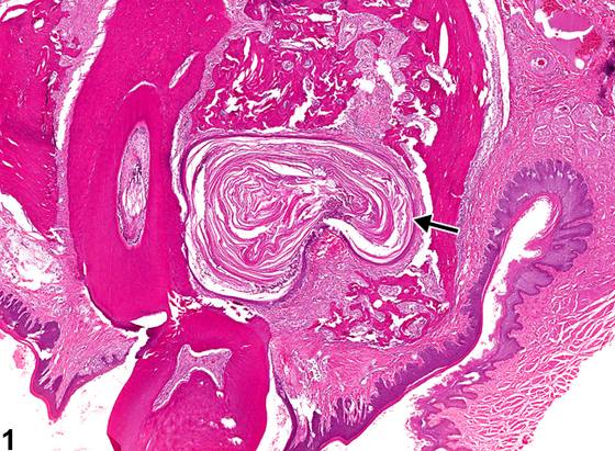
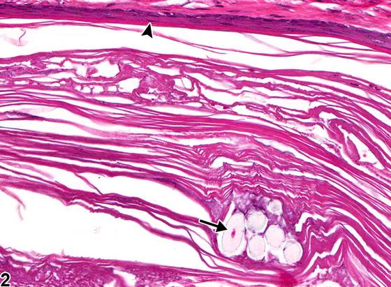
牙周袋(periodontal pocket)(Figure 1、Figure 2、Figure 3、Figure 4、Figure 5、Figure 6、Figure 7、Figure 8)是啮齿类动物常见的背景性病变。它们见于牙齿周围的牙周组织,尤其是磨牙周围。牙周袋表现为异常加深的牙龈沟,其内衬为角化的鳞状上皮,且常继发于毛发(Figure 1、Figure 2、Figure 3、Figure 4、Figure 5、Figure 6)、饲料(Figure 7、Figure 8)或垫料的嵌塞。另外,细菌性牙周炎可导致牙周膜纤维的脱离和吸收,并形成牙周袋,异物可在其中被包裹。嵌塞的毛发、饲料或垫料可穿透周围组织,导致慢性炎症和纤维化(Figure 4、Figure 5、Figure 6)。牙周袋内衬为鳞状上皮,该上皮常发生角化。牙周袋的鉴别诊断包括鳞状囊肿和囊性角化增生。囊性角化增生被认为处于从增生到鳞状细胞癌的连续谱上。相比之下,牙周袋往往是由正常鳞状上皮衬覆的单房结构。鳞状囊肿在形态学上与牙周袋相似,但它们不一定位于牙齿邻近部位,且不与口腔黏膜相通。理想情况下,在牙周袋病变中可以看到与口腔的相通,但由于切面原因,开口可能不明显。
诊断与分级建议(NTP 共识)
牙周袋应当诊断,但不进行分级。牙周袋内的异物(如毛干、垫料、饲料)不应单独诊断,而应在病理描述中加以描述。然而,在牙周袋周围组织内发现的异物应记录为“异物”。相关的炎症和纤维化不应单独诊断,除非其严重程度足以需要单独诊断。
图谱
英文原文
展开 NTP 官方英文描述与建议
Comment:
Periodontal pockets ( Figure 1 , Figure 2 , Figure 3 , Figure 4 , Figure 5 , Figure 6 , Figure 7 , and Figure 8 ) are common background findings in rodents. They are found in the periodontal tissues surrounding the teeth, especially the molars. The pockets represent an abnormally deepened gingival sulcus that is lined by keratinized squamous epithelium and are frequently secondary to the impaction of hair ( Figure 1 , Figure 2 , Figure 3 , Figure 4 , Figure 5 , and Figure 6 ), feed ( Figure 7 and Figure 8 ), or bedding material. Alternatively, bacterial periodontitis can result in detachment and resorption of periodontal ligament fibers and the formation of a periodontal pocket in which foreign bodies can become entrapped. Impacted hair, feed, or bedding may penetrate the surrounding tissues, resulting in chronic inflammation and fibrosis ( Figure 4 , Figure 5 , and Figure 6 ). They are lined by squamous epithelium that is often keratinized. Differential diagnoses for periodontal pocket include squamous cyst and cystic keratinizing hyperplasia. Cystic keratinizing hyperplasia is thought to be on a continuum from hyperplasia to squamous cell carcinoma. In comparison, periodontal pockets tend to be unilocular structures lined by normal squamous epithelium. Squamous cysts are similar to periodontal pockets morphologically, but they are not necessarily located adjacent to a tooth and do not communicate with the oral mucosa. Ideally, the communication to the oral cavity can be seen in a periodontal pocket lesion, but the opening may not be apparent because of the plane of section.
Recommendation:
Periodontal pockets should be diagnosed but not graded. Foreign material (e.g., hair shafts, bedding, feed) within the pocket should not be diagnosed separately but should be described in the pathology narrative. However, foreign material found in the tissue surrounding the pocket should be recorded as "foreign body." Associated inflammation and fibrosis should not be diagnosed separately unless warranted by severity.
来源与引用
原始条目:https://ntp.niehs.nih.gov/atlas/nnl/alimentary-system/tooth/PeriodontalPocket
图谱总目录:NTP Nonneoplastic Lesion Atlas。 内容属美国联邦政府作品(公有领域),可引用;引用时请标注来源。
建议引用格式:NTP Nonneoplastic Lesion Atlas. Periodontal Pocket (tooth). LabSmart 中文版:
https://www.labsmart.cn/ntp/tooth-periodontal-pocket.html
中文图谱总页(可展开对照全部 595 条):https://www.labsmart.cn/ntp-atlas.html#tooth-periodontal-pocket