牙髓骨性牙本质Pulp Osteodentin · 牙齿 · 消化系统 · NTP ntp-0259
诊断要点:骨样牙本质应根据骨样牙本质形成区域的数量和大小进行诊断和分级。牙齿或成牙本质细胞的任何损伤(若存在)应单独诊断和分级,并应在病理学描述中明确两者之间的关联。
病变描述
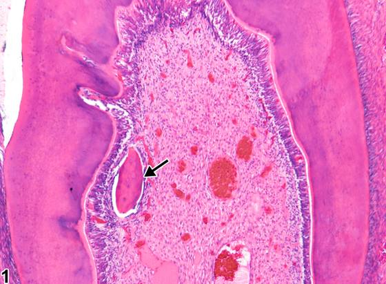
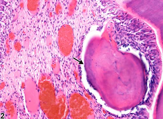
骨样牙本质(osteodentin)在牙髓受损或成牙本质细胞移位后形成。若牙齿发生折断,髓腔会被这种物质填充以将其封闭。此类反应也可作为一种继发性修复反应发生,以增加生长/萌出发生改变或中断的牙齿和/或处于改变了的生物力学应力下的牙齿的强度。骨样牙本质沉积的速率取决于刺激的强度和持续时间;刺激/损伤越严重,骨样牙本质沉积的速率越快。刺激的类型对骨样牙本质的结构和构造有影响。若刺激为龋损,由于细菌代谢产物和毒素的弥散,牙本质发生广泛破坏,牙髓受到损伤。因此,骨样牙本质快速沉积,呈稀疏而不规则的管状模式并伴有一些细胞包涵物。快速沉积后,成牙本质细胞被困于新形成的基质中,管状模式变得模糊或扭曲。形成可为孤立性(Figure 1、Figure 2,箭头)、多发性或弥漫性(Figure 3、Figure 4,箭头)。某些受试物可直接或间接引起细胞丰富区内的未分化牙髓间充质细胞终末分化为成牙本质细胞样细胞,并在牙髓内沉积骨样牙本质。骨样牙本质沉积物在髓腔内可类似于骨、骨样组织、前牙本质或牙本质。
诊断与分级建议(NTP 共识)
骨样牙本质应根据骨样牙本质形成区域的数量和大小进行诊断和分级。牙齿或成牙本质细胞的任何损伤(若存在)应单独诊断和分级,并应在病理学描述中明确两者之间的关联。
图谱
英文原文
展开 NTP 官方英文描述与建议
Comment:
Osteodentin is formed after injury to the pulp or displacement of odontoblasts. If a tooth fractures, the pulp chamber will fill with this material in order to seal it off. This type of reaction can also occur as a secondary reparative response to add strength to teeth with altered or disrupted growth/eruption and/or teeth under altered biomechanical stress. The rate of osteodentin deposition depends on the intensity and duration of the stimulus; the more severe the stimulus/injury, the more rapid the rate of osteodentin deposition. The type of stimulus has an effect on the architecture and structure of the osteodentin. If the stimulus is a carious lesion, there is extensive destruction of dentin and damage to the pulp, due to the differentiation of bacterial metabolites and toxins. Thus, osteodentin is deposited rapidly, with a sparse and irregular tubular pattern and some cellular inclusions. After rapid deposition, odontoblasts become trapped in the newly formed matrix, and the tubular pattern becomes obscured or distorted. Formation may be solitary ( Figure 1 and Figure 2 , arrows), multiple, or diffuse ( Figure 3 and Figure 4 , arrows). Some test articles can directly or indirectly cause undifferentiated pulp mesenchymal cells within the cell-rich zone to terminally differentiate into odontoblast-like cells and deposit osteodentin within the pulp. Osteodentin deposits may resemble bone, osteoid, predentin, or dentin in the pulp cavity.
Recommendation:
Osteodentin should be diagnosed and graded based on the number and size of areas of osteodentin formation. Any damage to the tooth or odontoblasts, if present, should be diagnosed and graded separately, and the association between the two should be made clear in the pathology narrative.
来源与引用
原始条目:https://ntp.niehs.nih.gov/atlas/nnl/alimentary-system/tooth/Pulp-Osteodentin
图谱总目录:NTP Nonneoplastic Lesion Atlas。 内容属美国联邦政府作品(公有领域),可引用;引用时请标注来源。
建议引用格式:NTP Nonneoplastic Lesion Atlas. Pulp Osteodentin (tooth). LabSmart 中文版:
https://www.labsmart.cn/ntp/tooth-pulp-osteodentin.html
中文图谱总页(可展开对照全部 595 条):https://www.labsmart.cn/ntp-atlas.html#tooth-pulp-osteodentin