肾上腺皮质副结节Accessory Adrenocortical Nodule · 肾上腺 · 内分泌系统 · NTP ntp-0085
诊断要点:肾上腺 - 副肾上腺皮质结节:无论何时出现均应诊断,但无需进行严重程度分级。若结节位于肾上腺周围脂肪(与肾上腺分离),则该组织应列为肾上腺周围脂肪。
病变描述
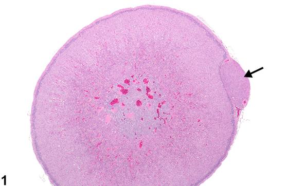
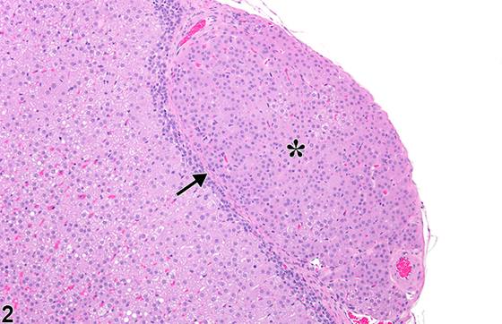
副肾上腺皮质结节(Accessory adrenocortical nodule)(Figure 1、Figure 2、Figure 3、Figure 4、Figure 5、Figure 6)在大鼠和小鼠中相当常见。该病变还有许多其他名称(如异位肾上腺组织、副皮质组织、错构瘤、副肾上腺、多余肾上腺和肾上腺皮质残留),副肾上腺皮质结节是起源于体腔上皮原基(肾上腺皮质即由此发生)部分或完全分离的残留物的轻微发育异常。它们最常位于与肾上腺一极被膜接触处(Figure 1、Figure 2、Figure 3、Figure 4),但也可完全与肾上腺分离,发生于腹膜后脂肪(Figure 5、Figure 6)、附着于肾被膜或腹膜腔任何部位。就其本身而言,副肾上腺皮质结节几乎总是无害的偶然发现,无临床或毒理学意义。然而,它们可发生与肾上腺皮质本身相同的退行性、增生性甚至肿瘤性改变。大多数结节为球形至卵圆形结构,周围通常有纤细的纤维性被膜,由分化良好的球状带型和束状带型细胞组成(Figure 1、Figure 2),有时混合存在,有时呈典型的分层排列。部分肾上腺皮质副组织由较疏松、外观更原始的皮质细胞巢组成(Figure 3、Figure 4)。典型的周围纤维性被膜可将副肾上腺皮质结节与局灶性(focal)皮质增生及皮质组织被膜外突出相区分。副结节还必须与皮质腺瘤和癌相鉴别,后者表现为细胞多形性和异型性及皮质分层丧失,但通常无被膜。
诊断与分级建议(NTP 共识)
肾上腺 - 副肾上腺皮质结节:无论何时出现均应诊断,但无需进行严重程度分级。若结节位于肾上腺周围脂肪(与肾上腺分离),则该组织应列为肾上腺周围脂肪。若结节与另一器官(如肾)相关,则该器官的异位组织是更合适的诊断(如肾 - 异位组织,肾上腺)。
图谱
英文原文
展开 NTP 官方英文描述与建议
Comment:
Accessory adrenocortical nodules ( Figure 1 , Figure 2 , Figure 3 , Figure 4 , Figure 5 , and Figure 6 ) are fairly common in rats and mice. Also known by many other terms (e.g., ectopic adrenal tissue, accessory cortical tissue, hamartoma, accessory adrenal gland, supernumary adrenal, and adrenocortical rest), accessory adrenocortical nodules are minor developmental anomalies arising from partly or completely detached rests of the coelomic epithelial primordia from which the adrenal cortex is derived. They are most frequently located in contact with the capsule at one pole of the adrenal gland ( Figure 1 , Figure 2 , Figure 3 , and Figure 4 ) but can occur completely separated from the adrenal gland in the retroperitoneal fat ( Figure 5 and Figure 6 ), attached to the kidney capsule, or anywhere in the peritoneal cavity. In and of themselves, accessory adrenocortical nodules are almost always innocuous incidental findings without clinical or toxicologic relevance. However, they can develop the same degenerative, hyperplastic, and even neoplastic changes as the adrenal cortex proper. Most nodules are spherical to ovoid structures surrounded by a usually delicate fibrous capsule and composed of well-differentiated zonae glomerulosa-type and fasciculata-type cells ( Figure 1 and Figure 2 ), sometimes admixed and sometimes arranged in typical layers. Some adrenocortical accessory tissue consists of less densely packed nests of more primitive-appearing cortical cells ( Figure 3 and Figure 4 ). The typical surrounding fibrous capsule distinguishes accessory adrenocortical nodules from focal cortical hyperplasia and from extracapsular protrusions of cortical tissue. Accessory nodules must also be distinguished from cortical adenomas and carcinomas, which exhibit cellular pleomorphism and atypia and loss of cortical layering but are not commonly encapsulated.
Recommendation:
Adrenal gland - Accessory adrenocortical nodule should be diagnosed whenever present, but a severity grade is not necessary. If a nodule is located in the periadrenal fat (separated from the adrenal gland), the tissue should be listed as periadrenal fat. If a nodule is associated with another organ (e.g., kidney), ectopic tissue of that organ is a more appropriate diagnosis (e.g., Kidney - Ectopic tissue, Adrenal gland).
来源与引用
原始条目:https://ntp.niehs.nih.gov/atlas/nnl/endocrine-system/adrenal-gland/AccessoryAdrenocorticalNodule
图谱总目录:NTP Nonneoplastic Lesion Atlas。 内容属美国联邦政府作品(公有领域),可引用;引用时请标注来源。
建议引用格式:NTP Nonneoplastic Lesion Atlas. Accessory Adrenocortical Nodule (adrenal-gland). LabSmart 中文版:
https://www.labsmart.cn/ntp/adrenal-gland-accessory-adrenocortical-nodule.html
中文图谱总页(可展开对照全部 595 条):https://www.labsmart.cn/ntp-atlas.html#adrenal-gland-accessory-adrenocortical-nodule