血管扩张Angiectasis · 肾上腺 · 内分泌系统 · NTP ntp-0087
诊断要点:肾上腺血管扩张应予以诊断并赋予严重程度分级。诊断中应包含部位修饰词(即cortex或medulla)以指示病变位置。
病变描述
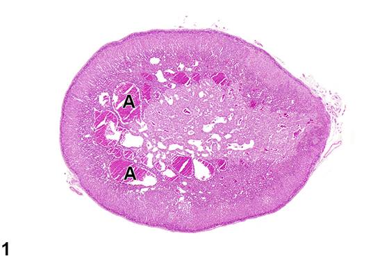
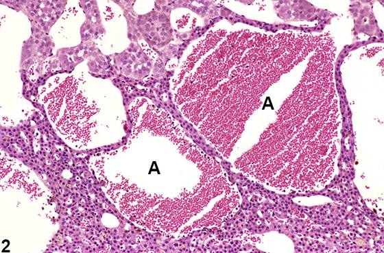
大鼠和小鼠肾上腺皮质和髓质的血管扩张(angiectasis)(Figure 1、Figure 2)以血管(毛细血管和/或静脉)的局灶性(focal)至弥漫性(diffuse)扩张为特征。扩张的血管排列疏松至不规则,不形成离散的团块。血管内衬单层分化良好的内皮细胞,由纤细的纤维性间质支撑,内含数量不等的血液。血管扩张可与其他肾上腺病变(如炎症、萎缩、变性和肿瘤)相关或继发于这些病变。它也可作为老年大鼠和小鼠的spontaneous年龄相关变化出现。血管扩张与血管瘤可能难以区分,但应尝试进行鉴别。血管瘤往往为边界清楚、无包膜的团块,由密集排列的扩张血管腔隙构成。血管腔隙内衬单层外观正常的内皮细胞,这些细胞排列在通常纤细的胶原性间隔上,尽管部分病例表现出更丰富的胶原性间质。相比之下,尽管血管扩张同样以扩张的血管通道为特征且内衬无明显异常的内皮,但血管通道排列更为疏松或不规则,而非表现为边界清楚的团块。
诊断与分级建议(NTP 共识)
肾上腺血管扩张应予以诊断并赋予严重程度分级。诊断中应包含部位修饰词(即cortex或medulla)以指示病变位置。如果皮质和髓质均存在,可省略部位修饰词并在病理学描述中描述位置。如果血管扩张被认为是其他病变(如囊性变性、炎症或肿瘤)的组成部分或继发于这些病变,则不应单独诊断,而应在病理学描述中予以描述。如果双侧肾上腺均可见血管扩张,诊断中应添加修饰词“双侧(bilateral)”(除非另有说明,病变假定为单侧)。
图谱
英文原文
展开 NTP 官方英文描述与建议
Comment:
Angiectasis ( Figure 1 and Figure 2 ) in the adrenal cortex and medulla of rats and mice is characterized by focal to diffuse dilation of blood vessels (capillaries and/or veins). The dilated vessels are loosely to irregularly arranged and do not form discrete masses. They are lined by single layers of well-differentiated endothelial cells supported by a delicate fibrous stroma and contain variable amounts of blood. Angiectasis can be associated with or secondary to other adrenal lesions such as inflammation, atrophy, degeneration, and neoplasia. It can also occur as a spontaneous age-related change in old rats and mice. Angiectasis and hemangioma can be difficult to differentiate, but a distinction should be attempted. Hemangiomas tend to be well-circumscribed, unencapsulated masses composed of densely packed, dilated vascular spaces. The vascular spaces are lined by a single layer of normal-appearing endothelial cells aligned on usually delicate collagenous septa, though some cases exhibit more abundant collagenous stroma. In contrast, although angiectasis is also characterized by dilated vascular channels lined by unremarkable endothelium, the vascular channels are more loosely or irregularly arranged rather than presenting as a well-demarcated mass.
Recommendation:
Adrenal angiectasis should be diagnosed and assigned a severity grade. A site modifier (i.e., cortex or medulla) should be included in the diagnosis to indicate the location of the lesion. If it is present in both the cortex and medulla, the site modifier may be omitted and the location described in the pathology narrative. If angiectasis is considered a component of or secondary to other lesions (e.g., cystic degeneration, inflammation, or neoplasia), it should not be diagnosed separately but should be described in the pathology narrative. If angiectasis is seen in both adrenal glands, the modifier "bilateral" should be added to the diagnosis (lesions are assumed to be unilateral unless otherwise indicated).
来源与引用
原始条目:https://ntp.niehs.nih.gov/atlas/nnl/endocrine-system/adrenal-gland/Angiectasis
图谱总目录:NTP Nonneoplastic Lesion Atlas。 内容属美国联邦政府作品(公有领域),可引用;引用时请标注来源。
建议引用格式:NTP Nonneoplastic Lesion Atlas. Angiectasis (adrenal-gland). LabSmart 中文版:
https://www.labsmart.cn/ntp/adrenal-gland-angiectasis.html
中文图谱总页(可展开对照全部 595 条):https://www.labsmart.cn/ntp-atlas.html#adrenal-gland-angiectasis