皮质萎缩Cortex Atrophy · 肾上腺 · 内分泌系统 · NTP ntp-0081
诊断要点:肾上腺皮质萎缩应当被诊断,并赋予严重程度分级和分布修饰词(即局灶性(focal)、弥漫性(diffuse))。
病变描述
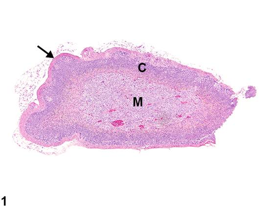
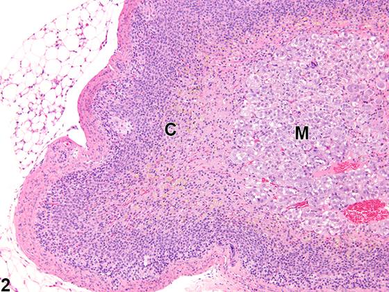
肾上腺皮质萎缩(Figure 1、Figure 2)可为局灶性(focal)或弥漫性(diffuse)。与正常肾上腺皮质(Figure 3、Figure 4)相比,萎缩的皮质以一层或多层皮质层厚度减少为特征,其原因是细胞体积减小或细胞丢失。束状带和网状带比球状带更常受累。腺体整体大小有不同程度的减小,常伴有腺体轮廓的扭曲。腺体被膜可因纤维化而增厚(Figure 1、Figure 2)。大鼠和小鼠肾上腺皮质萎缩的原因包括任何使糖皮质激素水平升高的因素,例如外源性给药或同侧或对侧腺体功能性肾上腺皮质肿瘤的内源性过度分泌,其继发性导致促肾上腺皮质激素(ACTH)水平降低。萎缩也可由更为直接的ACTH缺乏引起,例如可能随垂体功能异常而发生。皮质萎缩还可由外源性毒物直接作用引起,这些毒物干扰正常的肾上腺皮质类固醇生成和/或肾素-血管紧张素系统对肾上腺的生理作用。由于下丘脑、垂体、甲状腺和性腺与肾上腺之间复杂的生理相互作用,其他直接损伤这些组织或调节其分泌功能的外源性毒物和实验操作可在肾上腺皮质引起继发性效应,包括萎缩。皮质萎缩还可能是其他病理过程(如变性或坏死)的后遗症,也可与细胞质空泡化或色素蓄积同时存在。
诊断与分级建议(NTP 共识)
肾上腺皮质萎缩应当被诊断,并赋予严重程度分级和分布修饰词(即局灶性(focal)、弥漫性(diffuse))。相关病变(例如皮质变性或被膜纤维化)不应单独诊断,除非其严重程度足以支持单独诊断。如果双侧肾上腺均可见萎缩,应在诊断中加上修饰词“双侧(bilateral)”(除非另有说明,病变假定为单侧)。
图谱
英文原文
展开 NTP 官方英文描述与建议
Comment:
Adrenal cortical atrophy ( Figure 1 and Figure 2 ) may be focal or diffuse. Compared with the normal adrenal cortex ( Figure 3 and Figure 4 ), the atrophic cortex is characterized by reduced thickness of the one or more of the cortical layers due to a decrease in cell size or a loss of cells. The zonae fasciculata and reticularis are more often affected than the zona glomerulosa. There is variably decreased overall size of the gland, often with distortion of the gland outline. The glandular capsule may be thickened due to fibrosis ( Figure 1 and Figure 2 ). Causes of adrenal cortical atrophy in rats and mice include any factor that increases levels of glucocorticoids, such as exogenous administration or endogenous hypersecretion by a functional adrenocortical neoplasm in the same or contralateral gland, which secondarily results in decreased adrenocorticotropic hormone (ACTH) levels. Atrophy can also result from more direct ACTH deficiency, such as may occur with abnormal pituitary function. Cortical atrophy can also be a direct effect of exogenous toxicants that interfere with normal adrenocortical steroidogenesis and/or the physiologic effects of the renin-angiotensin system on the adrenal. Because of the complex physiologic interactions of hypothalamus, pituitary, thyroid, and gonads with the adrenal gland, other exogenous toxicants and experimental manipulations that directly damage these tissues or modulate their secretory functions can result in secondary effects in the adrenal cortex, including atrophy. Cortical atrophy may also be a sequela to other pathologic processes, such as degeneration or necrosis, and may also be concurrent with cytoplasmic vacuolization or pigment accumulation.
Recommendation:
Adrenal cortical atrophy should be diagnosed and assigned a severity grade and distribution modifier (i.e., focal, diffuse). Associated lesions (e.g., cortical degeneration or capsular fibrosis) should not be diagnosed separately unless warranted by severity. If atrophy is seen in both adrenal glands, the modifier "bilateral" should be added to the diagnosis (lesions are assumed to be unilateral unless otherwise indicated).
来源与引用
原始条目:https://ntp.niehs.nih.gov/atlas/nnl/endocrine-system/adrenal-gland/Cortex-Atrophy
图谱总目录:NTP Nonneoplastic Lesion Atlas。 内容属美国联邦政府作品(公有领域),可引用;引用时请标注来源。
建议引用格式:NTP Nonneoplastic Lesion Atlas. Cortex Atrophy (adrenal-gland). LabSmart 中文版:
https://www.labsmart.cn/ntp/adrenal-gland-cortex-atrophy.html
中文图谱总页(可展开对照全部 595 条):https://www.labsmart.cn/ntp-atlas.html#adrenal-gland-cortex-atrophy