皮质囊性变性Cortex Degeneration Cystic · 肾上腺 · 内分泌系统 · NTP ntp-0077
诊断要点:肾上腺皮质的囊性变性应当诊断并赋予严重程度分级。发生于增生或肥大灶内或皮质肿瘤内的囊性变性区域不应单独诊断,而应在病理学描述中加以描述。
病变描述
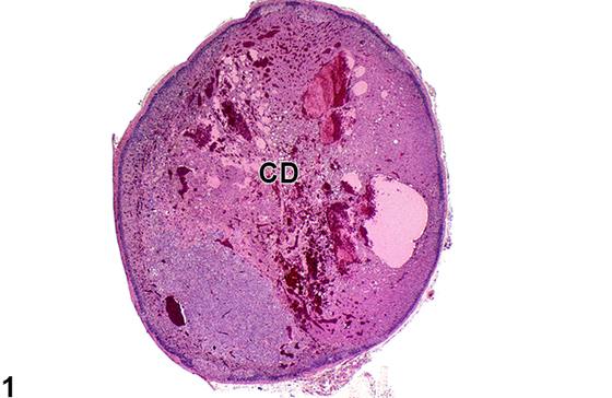
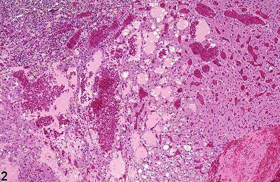
肾上腺皮质的囊性变性(cystic degeneration)(Figure 1、Figure 2、Figure 3、Figure 4、Figure 5)发生于许多品系的大鼠,但显然尚未在小鼠中报道。发生率随大鼠品系而异,某些品系(如 Sprague-Dawley、Wistar)的发生率远高于其他品系(如 F344)。在所有大鼠品系中,囊性变性在雌性中远比雄性常见。它通常是自发性的、与年龄相关的变化,但已有报道称给予某些化学物后发生率呈处理相关性的升高。囊性变性的早期阶段以束状带内小的、无压迫性的皮质窦隙增宽灶和血管扩张灶为特征;其间索内的皮质细胞可呈空泡化或轻度扁平。随着病变进展,受累区域扩大并可压迫邻近实质(Figure 3、Figure 4)。窦隙和血管扩张逐渐变得更加广泛,形成大的、常为融合性的囊样腔隙(Figure 1、Figure 2、Figure 3、Figure 4、Figure 5),其内充满蛋白性液体、红细胞和/或纤维素性血栓,但缺乏真性囊肿所特有的上皮衬里。空泡化或肿胀的皮质细胞团簇可散布于扩张的腔隙之间。囊性变性区域也可为大的皮质增生性或肥大性灶或皮质肿瘤的特征之一;然而,在囊性变性中,细胞数量较正常皮质减少。
诊断与分级建议(NTP 共识)
肾上腺皮质的囊性变性应当诊断并赋予严重程度分级。发生于增生或肥大灶内或皮质肿瘤内的囊性变性区域不应单独诊断,而应在病理学描述中加以描述。如果双侧肾上腺均可见囊性变性,则应在诊断中添加修饰词“双侧(bilateral)”(除非另有说明,病变假定为单侧)。
图谱
英文原文
展开 NTP 官方英文描述与建议
Comment:
Cystic degeneration of the adrenal cortex ( Figure 1 , Figure 2 , Figure 3 , Figure 4 , and Figure 5 ) occurs in many strains of rats but has apparently not been reported in mice. Incidences vary with rat strain, with some strains (e.g., Sprague-Dawley, Wistar) exhibiting much higher incidences than others (e.g., F344). In all rat strains, cystic degeneration is much more common in females than in males. It is usually a spontaneous, age-related change, though treatment-related increased incidences have been reported after administration of certain chemicals. Early stages of cystic degeneration are characterized by small, noncompressive foci of widened cortical sinusoids and dilated blood vessels in the zona fasciculata; cortical cells in the intervening cords can be vacuolated or slightly flattened. As the lesion progresses, affected areas enlarge and can compress adjacent parenchyma ( Figure 3 and Figure 4 ). The sinusoidal and vascular ectasia progressively becomes more extensive, resulting in large, often confluent, cyst-like cavitations ( Figure 1 , Figure 2 , Figure 3 , Figure 4 , and Figure 5 ) that are filled with proteinaceous fluid, red blood cells, and/or fibrinous thrombi but lack the epithelial lining characteristic of true cysts. Clusters of vacuolated or swollen cortical cells can be scattered among the dilated spaces. Areas of cystic degeneration can also be features of large cortical hyperplastic or hypertrophic foci or of cortical neoplasms; however, in cystic degeneration the number of cells is decreased compared with normal cortex.
Recommendation:
Cystic degeneration of the adrenal cortex should be diagnosed and assigned a severity grade. Areas of cystic degeneration occurring in foci of hyperplasia or hypertrophy or in cortical neoplasms should not be diagnosed separately but should be described in the pathology narrative. If cystic degeneration is seen in both adrenal glands, the modifier "bilateral" should be added to the diagnosis (lesions are assumed to be unilateral unless otherwise indicated).
来源与引用
原始条目:https://ntp.niehs.nih.gov/atlas/nnl/endocrine-system/adrenal-gland/Cortex-DegenerationCystic
图谱总目录:NTP Nonneoplastic Lesion Atlas。 内容属美国联邦政府作品(公有领域),可引用;引用时请标注来源。
建议引用格式:NTP Nonneoplastic Lesion Atlas. Cortex Degeneration Cystic (adrenal-gland). LabSmart 中文版:
https://www.labsmart.cn/ntp/adrenal-gland-cortex-degeneration-cystic.html
中文图谱总页(可展开对照全部 595 条):https://www.labsmart.cn/ntp-atlas.html#adrenal-gland-cortex-degeneration-cystic