皮质肥大Cortex Hypertrophy · 肾上腺 · 内分泌系统 · NTP ntp-0080
诊断要点:肾上腺皮质肥大应予以诊断,并赋予严重程度分级和适当的分布修饰词(即局灶性(focal)、弥漫性(diffuse))。
病变描述
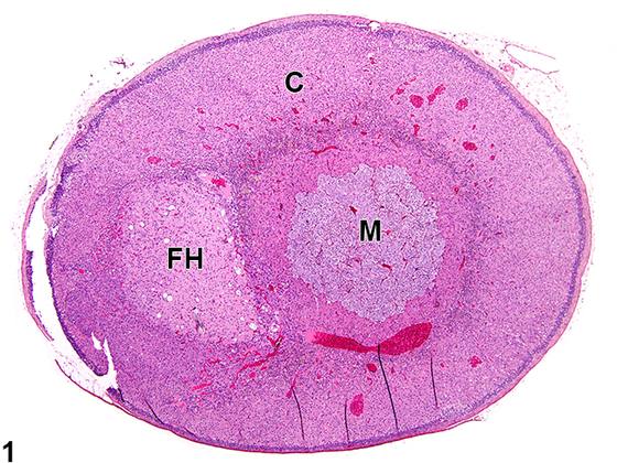
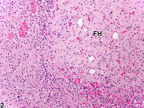
肾上腺皮质肥大(Adrenal cortical hypertrophy)是指细胞体积增大而细胞数量无明显增多。肥大的皮质细胞胞质增多,可呈淡染至亮嗜酸性(Figure 1、Figure 2、Figure 3、Figure 4),但也可呈透明、空泡化和/或嗜碱性。细胞核常增大。肥大灶内少数细胞核可具有非典型性特征。若肥大细胞中非典型细胞占较大比例,则应诊断为非典型性而非肥大(见Adrenal Gland - Cortex - Cellular Atypia)。皮质肥大可为局灶性(focal)或弥漫性(diffuse)。局灶性肥大(Figure 1、Figure 2、Figure 3、Figure 4)最常累及束状带。小的肥大灶通常无压迫性,但较大的局灶性病变可对邻近皮质实质造成不同程度的压迫(Figure 3)。与未受累的正常肾上腺(Figure 7、Figure 8)相比,弥漫性皮质肥大(Figure 5、Figure 6)常导致皮质明显增宽及腺体整体增大。大鼠和小鼠的(束状带)皮质肥大通常由促肾上腺皮质激素(adrenocorticotropic hormone,ACTH)水平升高所致,而ACTH升高又可源于多种原因,如原发性下丘脑或垂体疾病,或肾上腺皮质毒性或退行性病变所致的糖皮质激素反馈调节减弱。束状带的弥漫性、双侧(bilateral)且常为显著的肥大也可为各种原因所致应激的后遗症,该表现在大鼠中尤为常见。球状带细胞的肥大(及增生)可由肾素-血管紧张素系统紊乱导致血管紧张素II升高而引起。无论局灶性还是弥漫性,皮质肥大通常不被认为是肿瘤前性改变。然而,皮质肥大(细胞体积增大)和增生(细胞数量增多)常可同时存在于同一腺体甚至同一病灶中。因此,在实际操作中,确定某一病变和/或腺体中以哪种改变为主可能非常困难。皮质肥大还可与许多胞质空泡化病例中所见的皮质细胞增大相混淆。
诊断与分级建议(NTP 共识)
肾上腺皮质肥大应予以诊断,并赋予严重程度分级和适当的分布修饰词(即局灶性(focal)、弥漫性(diffuse))。当两侧腺体均存在肥大时,应使用修饰词“双侧(bilateral)”。应努力区分皮质肥大与增生,尽管这两种改变在同一病变中频繁叠加可使该判断非常具有挑战性。若同一病灶中两种所见同时存在,则仅记录增生,并在病理学描述中描述肥大特征。若肥大灶中存在少量非典型细胞,应在描述中予以描述。然而,若病理学家认为非典型细胞所占比例足够大,则应诊断细胞非典型性(见Adrenal Gland - Cortex - Cellular Atypia)。相关链接:Adrenal Gland - Cortex - Cellular Atypia
图谱
英文原文
展开 NTP 官方英文描述与建议
Comment:
Adrenal cortical hypertrophy refers to an increased cell size without an appreciable increase in cell numbers. Hypertrophic cortical cells have increased amounts of cytoplasm, which may be pale to brightly eosinophilic ( Figure 1 , Figure 2 , Figure 3 , and Figure 4 ) but may also be clear, vacuolated, and/or basophilic. The nucleus is frequently enlarged. Small numbers of nuclei in a focus of hypertrophy may have features of atypia. If a large proportion of the hypertrophic cells are atypical, then atypia should be diagnosed rather than hypertrophy (see Adrenal Gland - Cortex - Cellular Atypia ). Cortical hypertrophy may be focal or diffuse. Focal hypertrophy ( Figure 1 , Figure 2 , Figure 3 , and Figure 4 ) most often involves the zona fasciculata. Small hypertrophic foci are generally noncompressive, but larger focal lesions may cause variable compression of the adjacent cortical parenchyma ( Figure 3 ). Diffuse cortical hypertrophy ( Figure 5 and Figure 6 ) often results in pronounced widening of the cortex and overall gland enlargement compared with unaffected normal adrenal glands ( Figure 7 and Figure 8 ). Cortical hypertrophy (of the zona fasciculata) in rats and mice usually results from elevated levels of adrenocorticotropic hormone (ACTH), which in turn can be elevated due to various causes, such as primary hypothalamic or pituitary disease or decreased glucocorticoid feedback regulation caused by adrenal cortical toxic or degenerative lesions. Diffuse, bilateral, and often prominent hypertrophy in the zona fasciculata can also be a sequela to stress from various causes, a finding that is especially common in rats. Hypertrophy (and hyperplasia) of zona glomerulosa cells can result from derangements of the renin-angiotensin system that result in elevated angiotensin II. Whether focal or diffuse, cortical hypertrophy is generally not considered to be a preneoplastic change. However, cortical hypertrophy (increased cell size) and hyperplasia (increased cell numbers) can often be concurrent lesions in the same gland or even in the same focus. Thus, as a practical matter, determining which change is predominant in a given lesion and/or gland can be very difficult. Cortical hypertrophy can also be confused with the cortical cell enlargement that occurs in many cases of cytoplasmic vacuolization.
Recommendation:
Adrenal cortical hypertrophy should be diagnosed and assigned a severity grade and appropriate distribution modifier (i.e., focal, diffuse). The modifier "bilateral" should be used when hypertrophy is present in both glands. An attempt should be made to distinguish cortical hypertrophy from hyperplasia, though the frequent superimposition of both changes in the same lesion can make this determination very challenging. In cases where both findings are concurrent in the same focus, only hyperplasia should be recorded, with the hypertrophy feature described in the pathology narrative. If there are a small number of atypical cells in the focus of hypertrophy, they should be described in the narrative. However, if the pathologist feels there is a large enough proportion of atypical cells, cellular atypia should be diagnosed (see Adrenal Gland - Cortex - Cellular Atypia ). Related Links: Adrenal Gland - Cortex - Cellular Atypia
来源与引用
原始条目:https://ntp.niehs.nih.gov/atlas/nnl/endocrine-system/adrenal-gland/Cortex-Hypertrophy
图谱总目录:NTP Nonneoplastic Lesion Atlas。 内容属美国联邦政府作品(公有领域),可引用;引用时请标注来源。
建议引用格式:NTP Nonneoplastic Lesion Atlas. Cortex Hypertrophy (adrenal-gland). LabSmart 中文版:
https://www.labsmart.cn/ntp/adrenal-gland-cortex-hypertrophy.html
中文图谱总页(可展开对照全部 595 条):https://www.labsmart.cn/ntp-atlas.html#adrenal-gland-cortex-hypertrophy