皮质细胞质空泡化Cortex Vacuolization Cytoplasmic · 肾上腺 · 内分泌系统 · NTP ntp-0079
诊断要点:肾上腺皮质细胞质空泡化应予以诊断并赋予严重程度分级。诊断中还应包括适当的分布修饰词(即局灶性(focal)或弥漫性(diffuse))。
病变描述
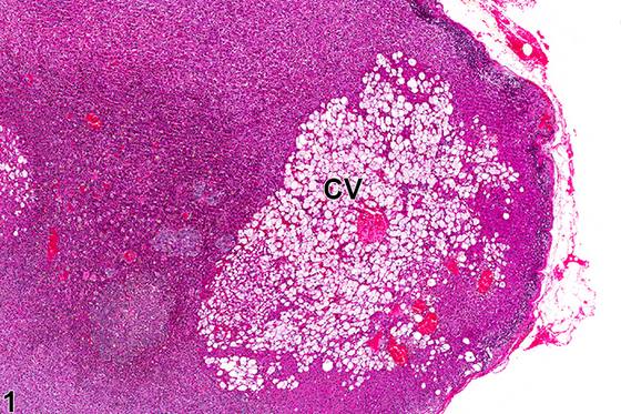
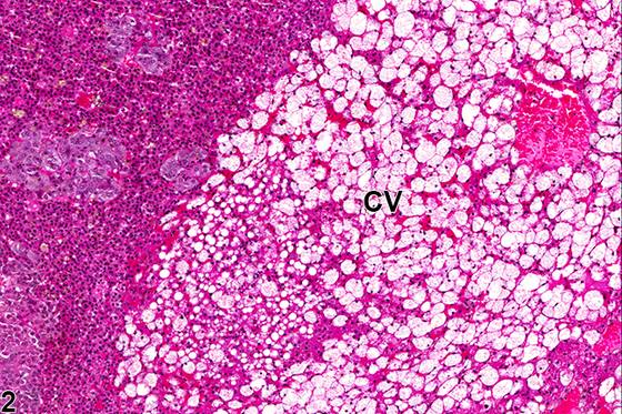
大鼠和小鼠的肾上腺皮质空泡化(adrenal cortical vacuolization)的特征是皮质细胞内积聚通常为清亮的空泡,主要位于束状带(zona fasciculata),尽管网状带(zona reticularis)和球状带(zona glomerulosa)也常受累。空泡化可为局灶性(focal)(Figure 1、Figure 2)或弥漫性(diffuse)(Figure 3、Figure 4)。即使较大的局灶性病变通常也几乎不引起压迫,或无压迫。空泡可为单个至几个大空泡,也可为多个较小的空泡。在许多情况下,已证明这些空泡代表各种脂质或胆固醇的积聚。皮质空泡化可作为自发性、年龄相关的变化(尤其在大鼠中)发生;继发于各种原因引起的应激;以及由给予许多外源性药物和毒物引起,尤其是那些干扰肾上腺皮质正常类固醇生成和/或扰乱下丘脑-垂体-肾上腺激素轴(包括促肾上腺皮质激素[ACTH]的正常分泌和代谢)的药物和毒物。皮质空泡化也可由实验性给予过量ACTH或皮质酮引起。
诊断与分级建议(NTP 共识)
肾上腺皮质细胞质空泡化应予以诊断并赋予严重程度分级。诊断中还应包括适当的分布修饰词(即局灶性(focal)或弥漫性(diffuse))。如果局限于皮质的某一特定带,应在病理学叙述中注明。在伴有部分组成性空泡化细胞的肾上腺皮质增生或肥大的病例中,空泡化不应单独诊断,但应在叙述中描述为增生或肥大的一个特征。如果两侧肾上腺均见空泡化,诊断中应加上修饰词“双侧(bilateral)”(除非另有说明,病变假定为单侧)。
图谱
英文原文
展开 NTP 官方英文描述与建议
Comment:
Adrenal cortical vacuolization in rats and mice is characterized by the accumulation of usually clear vacuoles within cortical cells, mainly in the zona fasciculata, although the zonae reticularis and glomerulosa are also often affected. The vacuolization can be focal ( Figure 1 and Figure 2 ) or diffuse ( Figure 3 and Figure 4 ). Even large focal lesions generally cause little to no compression. The vacuoles can range from a single to a few large vacuoles to multiple smaller ones. In many cases, the vacuoles have been shown to represent accumulations of various lipids or cholesterol. Cortical vacuolization can occur as a spontaneous, age-related change (especially in rats); secondary to stress from various causes; and from administration of many exogenous drugs and toxins, especially those that interfere with normal steroidogenesis in the adrenal cortex and/or perturb the hypothalamic-pituitary-adrenal hormonal axis (including normal secretion and metabolism of adrenocorticotropic hormone [ACTH]). Cortical vacuolization can also result from experimental administration of excess ACTH or corticosterone.
Recommendation:
Adrenal cortical cytoplasmic vacuolization should be diagnosed and assigned a severity grade. An appropriate distribution modifier (i.e., focal or diffuse) should also be included in the diagnosis. If localized to a particular zone of the cortex, this should be noted in the pathology narrative. In cases of adrenal cortical hyperplasia or hypertrophy with some constituent vacuolated cells, the vacuolization should not be diagnosed separately, though it should be described in the narrative as a feature of the hyperplasia or hypertrophy. If vacuolization is seen in both adrenal glands, the modifier "bilateral" should be added to the diagnosis (lesions are assumed to be unilateral unless otherwise indicated).
来源与引用
原始条目:https://ntp.niehs.nih.gov/atlas/nnl/endocrine-system/adrenal-gland/Cortex-VacuolizationCytoplasmic
图谱总目录:NTP Nonneoplastic Lesion Atlas。 内容属美国联邦政府作品(公有领域),可引用;引用时请标注来源。
建议引用格式:NTP Nonneoplastic Lesion Atlas. Cortex Vacuolization Cytoplasmic (adrenal-gland). LabSmart 中文版:
https://www.labsmart.cn/ntp/adrenal-gland-cortex-vacuolization-cytoplasmic.html
中文图谱总页(可展开对照全部 595 条):https://www.labsmart.cn/ntp-atlas.html#adrenal-gland-cortex-vacuolization-cytoplasmic