皮质X带萎缩Cortex X Zone Atrophy · 肾上腺 · 内分泌系统 · NTP ntp-0076
诊断要点:肾上腺皮质X带退化是正常生理过程,其各阶段常为雌雄两性及不同年龄小鼠的偶然发现。
病变描述
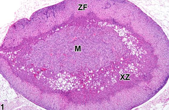
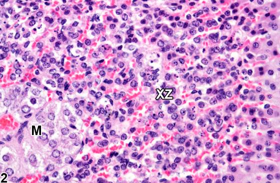
X带在小鼠出生后数天出现于雌雄两性,至断奶时充分发育。X带位于皮质与髓质交界处,其内细胞质较束状带细胞更具嗜酸性(Figure 1、Figure 2)。在雄性小鼠中,X带于青春期(约5周龄)退化。在雌性中,X带在青春期后持续数周,随后在未产雌性中较为逐渐地退化,或在首次妊娠时较为迅速地退化。X带的发育程度及退化速率也可随小鼠品系而异。在许多小鼠品系(包括B6C3F1品系)的雌性中,X带的正常退化(复旧)以形态学上不同的阶段进展。退化的起始以散在组成细胞的空泡化为开端(Figure 1、Figure 2)。随着退化继续,空泡化细胞的数量逐渐增加,直至几乎所有X带细胞均受累。在后期阶段,空泡化的X带细胞发生变性和坏死,随后整体X带结构崩解、凝缩并最终消失。常见的终末期后遗症为含色素细胞残留积聚于原先由X带占据的髓质周围区域。在雄性中,X带退化相似,只是通常不发生空泡化。X带的功能未知。其正常发育和退化由性腺激素和甲状腺激素介导,因此改变这些激素水平的因素可影响X带。例如,去势可延长雌性小鼠及青春期前雄性小鼠中X带的存留时间,并可导致青春期后雄性中X带重新出现。给予雄激素(如睾酮)后,雌性小鼠的X带迅速消失。给予某些其他化学物也可影响X带,导致非同步性偏差,例如与年龄匹配的同批对照(Figure 1、Figure 2)相比,处理组中出现加速退化(Figure 3、Figure 4)。
诊断与分级建议(NTP 共识)
肾上腺皮质X带退化是正常生理过程,其各阶段常为雌雄两性及不同年龄小鼠的偶然发现。正常X带退化阶段的特征(如空泡化、变性)及其终末期后遗症(如X带崩解、纤维化和色素细胞积聚)不应被误认为病理学病变。因此,在给定研究中,当生理性X带退化以相似的、与年龄和性别相适应的方式发生于对照和处理小鼠两者时,不应诊断该退化。异常加速(快速)退化,或相反地,X带过度长久存留,可为各种化学物和外源性激素处理所致效应。然而,在这些情况下,处理动物的X带形态将不具独特性,也不表现出特征性病理学特征。相反,毒性效应的存在仅表现为处理动物X带外观(时间阶段)与正常、与年龄和性别相适应的同批研究对照相比存在差异。由于这些原因,处理动物的X带毒性不能孤立地诊断,而必须在研究对照中所发生的生理性时间阶段的背景下进行评估。
图谱
英文原文
展开 NTP 官方英文描述与建议
Comment:
The X-zone appears a few days after birth in mice of both sexes and is fully developed by weaning. The X-zone is located at the junction of the cortex and the medulla and is populated by cells with more eosinophilic cytoplasm than those of the zona fasciculata ( Figure 1 and Figure 2 ). In male mice, the X-zone regresses at puberty (by about 5 weeks of age). In females, the X-zone persists for several weeks past puberty and then regresses more gradually in nulliparous females or more rapidly at first pregnancy. The extent of X-zone development and the rate of involution can also vary with mouse strain. Normal regression (involution) of the X-zone in females of many mouse strains, including the B6C3F1 strain, progresses in morphologically distinct stages. The onset of regression begins with vacuolization of scattered constituent cells ( Figure 1 and Figure 2 ). As regression continues, the number of vacuolated cells progressively increases until virtually all X-zone cells are affected. In later stages, the vacuolated X-zone cells undergo degeneration and necrosis, with subsequent overall X-zone architectural collapse, condensation, and eventual disappearance. A common end-stage sequela is the residual accumulation of pigment-laden cells in the perimedullary area formerly occupied by the X-zone. In males, X-zone regression is similar except that it usually occurs without vacuolization. The function of the X-zone is unknown. Its normal development and regression are mediated by gonadal and thyroid hormones, so factors that alter levels of these hormones can affect the X-zone. For example, gonadectomy prolongs the persistence of the X-zone in female mice and prepubertal male mice and can cause the reappearance of the X-zone in postpubertal males. Administration of androgens like testosterone is followed by rapid disappearance of the X-zone in female mice. Administration of certain other chemicals can also affect the X-zone, resulting in asynchronous deviations, such as accelerated regression ( Figure 3 and Figure 4 ) in treated groups compared with age-matched concurrent controls ( Figure 1 and Figure 2 ).
Recommendation:
Adrenal cortical X-zone regression is a normal physiologic process, and its various stages are often incidental findings in mice of both sexes and at various ages. Features of normal X-zone regression stages (e.g., vacuolization, degeneration) and its end-stage sequelae (e.g., X-zone collapse, fibrosis, and pigment-cell accumulation) should not be mistaken for pathologic lesions. Thus, physiologic X-zone regression should not be diagnosed when it occurs in a similar, age- and sex-appropriate manner in both the control and treated mice in a given study. Abnormally accelerated (rapid) regression or, conversely, excessively lengthy persistence of the X-zone can be effects of treatment with various chemicals and exogenous hormones. However, in these cases, the X-zone morphology of treated animals will not be distinctive or exhibit pathognomonic pathologic features. Instead, the existence of a toxic effect manifests only as a disparity in the appearance (temporal stage) of the X-zone in treated animals compared with normal, age- and sex-appropriate concurrent study controls. For these reasons, X-zone toxicity in treated animals cannot be diagnosed in isolation but must be evaluated in the context of the physiologic temporal stage occurring in the study controls.
来源与引用
原始条目:https://ntp.niehs.nih.gov/atlas/nnl/endocrine-system/adrenal-gland/CortexX-Zone-Atrophy
图谱总目录:NTP Nonneoplastic Lesion Atlas。 内容属美国联邦政府作品(公有领域),可引用;引用时请标注来源。
建议引用格式:NTP Nonneoplastic Lesion Atlas. Cortex X Zone Atrophy (adrenal-gland). LabSmart 中文版:
https://www.labsmart.cn/ntp/adrenal-gland-cortex-x-zone-atrophy.html
中文图谱总页(可展开对照全部 595 条):https://www.labsmart.cn/ntp-atlas.html#adrenal-gland-cortex-x-zone-atrophy