色素Pigment · 肾上腺 · 内分泌系统 · NTP ntp-0086
诊断要点:看似与处理相关原发改变的色素沉积应予以诊断,并赋予严重程度分级和部位修饰词(即皮质、髓质或被膜)。
病变描述
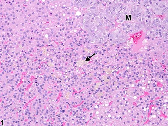
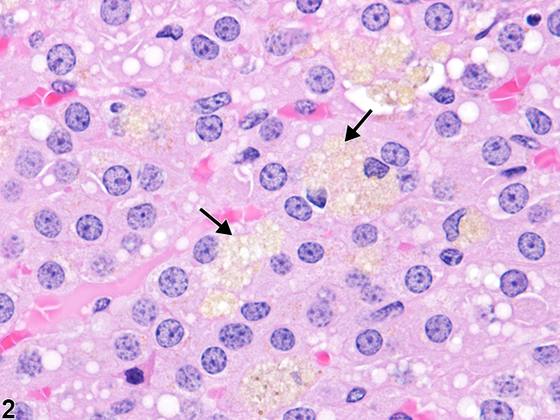
肾上腺中的色素最常见于皮质,但也可见于髓质和肾上腺被膜。小鼠和大鼠的肾上腺皮质色素通常为脂褐素沉积,其特征为胞质内黄色至深棕色、颗粒状至无定形物质,充盈并使肾上腺皮质细胞扩张。肿胀细胞的胞质可呈现不同程度的空泡化或“泡沫状”(Figure 1、Figure 2)。脂褐素沉积可具有自发荧光,通常为苏丹染色阳性,且过碘酸-雪夫(PAS)染色和抗酸染色阳性;铁染色阳性程度不一。含铁血黄素色素也可作为出血的后遗症而存在。含铁血黄素色素通常见于巨噬细胞,呈金黄色至深棕色。脂褐素是指胞质内、通常为溶酶体内、不可降解的脂质和蛋白质氧化/聚合残留物(以及其他部分,如晚期糖基化终产物)的异质性混合物,其因衰老或其他因素而在各种体细胞中蓄积。过度和/或时间上不适当的肾上腺脂褐素色素聚集(与年龄匹配的对照相比)也可为皮质细胞变性和/或萎缩(由各种原因所致)的特征,或可由饮食失衡(如维生素E缺乏)或给予某些外源性化学物质所致。在小鼠中,充满脂褐素色素的肾上腺皮质细胞最常作为X带生理性退化的晚期特征或后遗症出现。因此,它们是老年小鼠中常见的偶然发现。充满色素的细胞聚集体主要位于毗邻皮髓质交界处、原为X带占据的区域。较轻、时间上进展较少的病例由散在的充满色素细胞的小灶组成,但随着动物年龄增长,细胞簇常增大并可融合成环绕髓质的环状带。含有类似脂褐素和/或含铁血黄素的巨噬细胞可散布于受累皮质细胞之间。在大鼠中,肾上腺皮质内含脂褐素色素的细胞也是一种常见的年龄相关偶然性改变。尽管大鼠没有X带,但充满色素的细胞往往最常见于内皮质,通常为单个细胞或非常小的细胞簇(Figure 1、Figure 2),不同于小鼠中常见的大型、显著的聚集体。
诊断与分级建议(NTP 共识)
看似与处理相关原发改变的色素沉积应予以诊断,并赋予严重程度分级和部位修饰词(即皮质、髓质或被膜)。如果同时存在于皮质和髓质,可省略部位修饰词,并在病理学叙述中描述其位置。在各种组织的组织学切片中,即使使用一系列特殊染色,明确鉴定色素也往往很困难。因此,推荐的诊断为“色素”,不加类型修饰词(而非诊断色素类型,例如“色素,含铁血黄素”或“色素,脂褐素”)。色素的形态学特征应在病理学叙述中描述。然而,并非所有色素沉积都必须诊断,因为有些几乎是老年动物中普遍存在的自发性改变和/或继发于无关的疾病过程。与小鼠X带退化相关的色素不应单独诊断。在大鼠和小鼠中作为自发性衰老改变而蓄积的皮质细胞色素不应诊断,除非在发生率或严重程度上存在处理相关差异。病理学家应自行判断此类继发性色素沉积是否显著到足以需要单独诊断。
图谱
英文原文
展开 NTP 官方英文描述与建议
Comment:
Pigments in the adrenal gland are most commonly found in the cortex but can also be seen in the medulla and adrenal capsule. Adrenal cortical pigment in mice and rats is usually lipofuscin deposition, which is characterized by the intracytoplasmic, yellow to dark brown, granular to amorphous material that fills and distends adrenal cortical cells. The cytoplasm of the swollen cells may appear variably vacuolated or "foamy" ( Figure 1 and Figure 2 ). Lipofuscin deposits may be autofluorescent and are usually sudanophilic and positive with periodic acid-Schiff (PAS) and acid-fast stains; positivity with iron stains is variable. Hemosiderin pigment may also be present as a sequela to hemorrhage. Hemosiderin pigment is typically found in macrophages and is golden brown to dark brown. Lipofuscin refers to heterogeneous mixtures of intracytoplasmic, usually intralysosomal, nondegradable residues of lipid and protein oxidation/polymerization (as well as other moieties, such as advanced glycosylated end products) that accumulate in various somatic cells due to aging or other factors. Excessive and/or temporally inappropriate adrenal lipofuscin pigment aggregations (compared with age-matched controls) can also be a feature of cortical cell degeneration and/or atrophy (due to various causes) or can result from dietary imbalances (e.g., vitamin E deficiency) or administration of certain exogenous chemicals. In mice, lipofuscin pigment-laden adrenal cortical cells occur most often as late-stage features or sequelae to physiologic regression of the X-zone. Thus, they are common incidental findings in aging mice. The pigment-laden cell aggregates are located primarily in the region adjacent to the corticomedullary junction formerly occupied by the X-zone. Milder, less temporally advanced cases consist of scattered small foci of pigment-laden cells, but as the animal ages the cell clusters often enlarge and can coalesce into circumferential bands surrounding the medulla. Macrophages containing similar lipofuscin and/or hemosiderin can be interspersed among the affected cortical cells. In rats, cells in the adrenal cortex containing lipofuscin pigment are also a common age-related incidental change. Even though rats do not have an X-zone, the pigment-laden cells tend to be most common in the inner cortex, usually as single cells or very small clusters ( Figure 1 and Figure 2 ), unlike the large, prominent aggregates often seen in mice.
Recommendation:
Pigment deposits that appear to be primary treatment-related changes should be diagnosed and assigned a severity grade and site modifier (i.e., cortex, medulla, or capsule). If it is present in both the cortex and medulla, the site modifier may be omitted and the location described in the pathology narrative. Definitive identification of pigments in various tissues is often difficult in histologic sections, even with a battery of special stains. Therefore, the recommended diagnosis is "pigment" without a type modifier (as opposed to diagnosing the type of pigment, e.g., "pigment, hemosiderin" or "pigment, lipofuscin"). The morphologic features of the pigment should be described in the pathology narrative. Not all pigment deposits have to be diagnosed, however, as some are almost ubiquitous spontaneous changes in aging animals and/or secondary to extraneous disease processes. Pigment associated with X-zone regression in mice should not be diagnosed separately. The cortical cell pigment that accumulates as a spontaneous aging change in rats and mice should not be diagnosed unless there are treatment-related differences in incidence or severity. The pathologist should use his or her judgment as to whether such secondary pigment deposits are prominent enough to warrant a separate diagnosis.
来源与引用
原始条目:https://ntp.niehs.nih.gov/atlas/nnl/endocrine-system/adrenal-gland/Pigment
图谱总目录:NTP Nonneoplastic Lesion Atlas。 内容属美国联邦政府作品(公有领域),可引用;引用时请标注来源。
建议引用格式:NTP Nonneoplastic Lesion Atlas. Pigment (adrenal-gland). LabSmart 中文版:
https://www.labsmart.cn/ntp/adrenal-gland-pigment.html
中文图谱总页(可展开对照全部 595 条):https://www.labsmart.cn/ntp-atlas.html#adrenal-gland-pigment