血管扩张Angiectasis · 骨髓 · 造血系统 · NTP ntp-0436
诊断要点:在亚慢性研究和慢性研究中,只要存在血管扩张,就应进行诊断并分级。与处理相关的血管扩张还应在病理学叙述中描述。
病变描述
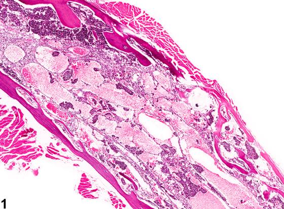
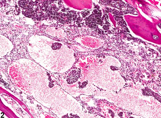
骨髓的血管供应最终以毛细血管开口于窦状隙丛而告终,窦状隙内衬内皮细胞,外层为不完整的外膜细胞层。骨髓血管扩张(angiectasis)定义为这些血管或窦状隙被血液扩张。在某些情况下,扩张可严重到出现大的红细胞和/或蛋白性液体(血清)湖,不应将其与出血混淆(Figure 1、Figure 2)。连续完整的内皮衬里的存在有助于将血管扩张与出血区分开来。随着血管系统更严重或慢性的充血,有时可在扩张的血管周围见到含有色素和/或胆固醇裂隙的巨噬细胞。应尝试区分血管扩张与血管瘤(hemangioma),尽管这种区分并不总是显而易见。血管瘤往往边界清楚、无包膜,由紧密排列的扩张血管腔隙构成。每个血管腔隙均由单层外观正常的内皮细胞围成并衬里,这些内皮细胞排列于胶原性间隔上,间隔通常较薄,尽管有些具有宽阔的胶原性间质。血管扩张通常不表现为边界清楚的肿块:扩张的血管通道常不规则地穿行于造血组织中。血管扩张已在造血组织严重丧失的啮齿动物中观察到。它也作为非特异性所见被观察到,或与炎症、肿瘤形成或血管/心血管疾病(例如血栓、充血性心力衰竭)相关。
诊断与分级建议(NTP 共识)
在亚慢性研究和慢性研究中,只要存在血管扩张,就应进行诊断并分级。与处理相关的血管扩张还应在病理学叙述中描述。分级基于扩张腔隙的数量和扩张程度的综合评估。不应使用术语“ectasia”代替血管扩张。
图谱
英文原文
展开 NTP 官方英文描述与建议
Comment:
The vascular supply of the bone marrow culminates with capillaries opening into a plexus of sinusoids lined with endothelial cells and an incomplete outer layer of adventitial cells. Angiectasis of the bone marrow is defined as dilatation of these vessels or sinusoids with blood. In some cases, the dilatation can be so severe as to cause the appearance of large lakes of erythrocytes and/or proteinaceous fluid (serum) that should not be confused with hemorrhage ( Figure 1 and Figure 2 ). The presence of a continuous intact endothelial lining helps distinguish angiectasis from hemorrhage. With more severe or chronic congestion of the vasculature, macrophages sometimes containing pigment and/or cholesterol clefts may be seen around the dilated vasculature. A distinction between angiectasis and hemangioma should be attempted, although the distinction is not always obvious. Hemangiomas tend to be well-circumscribed, unencapsulated masses composed of tightly packed, dilated vascular spaces. Each vascular space is enclosed and lined by a single layer of normal-appearing endothelial cells aligned on collagenous septa, which are usually thin, although some have broad collagenous stroma. Angiectasis does not usually present as a well-circumscribed mass: the dilated vascular channels often course irregularly through the hematopoietic tissue. Angiectasis has been observed in rodents with severe loss of hematopoietic tissue. It is also observed as a nonspecific finding or in association with inflammation, neoplasia, or vascular/cardiovascular disorders (e.g., thrombi, congestive heart failure).
Recommendation:
Angiectasis should be diagnosed and graded in subchronic and chronic studies whenever it is present. Treatment-related angiectasis should also be described in the pathology narrative. Grading is based on a combination of the number of dilated spaces and the degree of dilatation. The term “ectasia” should not be used in place of angiectasis.
来源与引用
原始条目:https://ntp.niehs.nih.gov/atlas/nnl/hematopoietic-system/bone-marrow/Angiectasis
图谱总目录:NTP Nonneoplastic Lesion Atlas。 内容属美国联邦政府作品(公有领域),可引用;引用时请标注来源。
建议引用格式:NTP Nonneoplastic Lesion Atlas. Angiectasis (bone-marrow). LabSmart 中文版:
https://www.labsmart.cn/ntp/bone-marrow-angiectasis.html
中文图谱总页(可展开对照全部 595 条):https://www.labsmart.cn/ntp-atlas.html#bone-marrow-angiectasis