增生Hyperplasia · 凝固腺 · 雄性生殖系统 · NTP ntp-0438
诊断要点:增生应进行分级并记录,当双侧凝固腺均受累时应注明为双侧性。严重程度应依据受累较重的腺体判定。
病变描述
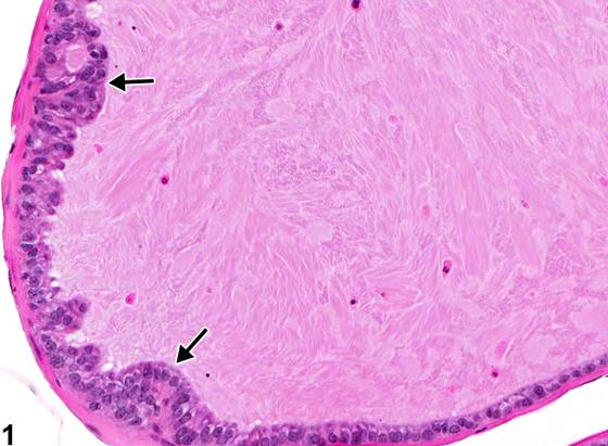
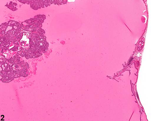
凝固腺增生由衬覆上皮的增殖构成。细胞可堆积(箭头,Figure 1)、形成短乳头状皱褶,或向腺泡腔内突出。Figure 1所示的增生为多灶性(multifocal)但轻微(mild);在Figure 2、Figure 3中则为focally extensive,并在腺泡腔内形成乳头状皱褶。凝固腺增生很可能是与年龄相关的改变。产前暴露于雌激素类物质(包括己烯雌酚)所致的雌性化,已在啮齿类动物中与凝固腺的增生性和化生性改变相关联。
诊断与分级建议(NTP 共识)
增生应进行分级并记录,当双侧凝固腺均受累时应注明为双侧性。严重程度应依据受累较重的腺体判定。与其他附属性腺中可能存在的类似改变的相关性,应在病理学描述中提及。
图谱
英文原文
展开 NTP 官方英文描述与建议
Comment:
Hyperplasia of the coagulating gland consists of proliferation of the lining epithelium. The cells may pile up (arrows, Figure 1 ), form short papillary folds, or bulge into the acinar lumen. Hyperplasia shown in Figure 1 is multifocal but mild; in Figure 2 and Figure 3 it is focally extensive, with formation of papillary folds in the acinar lumen. Hyperplasia of the coagulating gland is likely an age-related change. Feminization from prenatal exposure to estrogenic agents, including diethylstilbestrol, has been associated with hyperplastic and metaplastic changes in the coagulating glands of rodents.
Recommendation:
Hyperplasia should be graded and recorded, with qualification as bilateral when both coagulating glands are affected. Severity should be based on the more severely affected gland. Correlations with similar changes that may be present in other accessory sex gland should be mentioned in the pathology narrative.
来源与引用
原始条目:https://ntp.niehs.nih.gov/atlas/nnl/reproductive-system-male/coagulating-gland/Hyperplasia
图谱总目录:NTP Nonneoplastic Lesion Atlas。 内容属美国联邦政府作品(公有领域),可引用;引用时请标注来源。
建议引用格式:NTP Nonneoplastic Lesion Atlas. Hyperplasia (coagulating-gland). LabSmart 中文版:
https://www.labsmart.cn/ntp/coagulating-gland-hyperplasia.html
中文图谱总页(可展开对照全部 595 条):https://www.labsmart.cn/ntp-atlas.html#coagulating-gland-hyperplasia