上皮空泡化Epithelium Vacuolation · 附睾 · 雄性生殖系统 · NTP ntp-0495
诊断要点:上皮的空泡化应记录并分级,并且如果其发生率和/或严重程度似乎与化学物给药相关,则应在病理学叙述中加以讨论。
病变描述
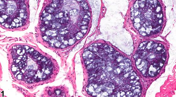
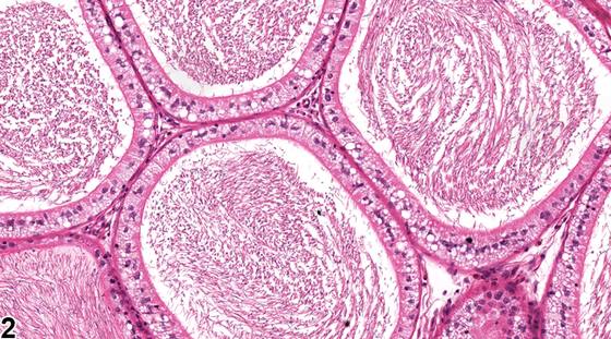
附睾上皮可发生多种不同形式的空泡化。这些空泡化常呈节段性和区域特异性。空泡化可能是多种退行性过程的结果,包括脂质、磷脂(磷脂沉积症)、糖蛋白或液体的蓄积。空泡可以较大(大空泡化,Figure 1)或较小(小空泡化,Figure 2),并可表现为空虚或充满嗜碱性或嗜酸性物质。嗜碱性空泡化是大鼠附睾头远端特定节段常见的年龄相关背景性所见(Figure 1)。大的透明空泡常作为偶然性所见见于大鼠附睾体尾交界处。附睾空泡化也可与受试物相关,并可呈弥漫性或节段性。透明细胞是附睾上皮的正常细胞类型,参与腔内颗粒物质的胞吞作用。当附睾腔内有细胞碎片增多时,透明细胞的数量和显著程度常增加,尤其是在附睾尾(Figure 3)。应注意将这些正常透明细胞与退行性空泡性改变相区分。由于透明细胞充满溶酶体,而溶酶体经Periodic acid-Schiff染色呈强阳性,因此可作出区分。
诊断与分级建议(NTP 共识)
上皮的空泡化应记录并分级,并且如果其发生率和/或严重程度似乎与化学物给药相关,则应在病理学叙述中加以讨论。如果空泡化的类型和位置具有特异性且值得注意,则应予以注明。诊断应指明该改变是否为双侧性。建议与其他雄性生殖器官的紊乱进行关联分析,以助于解释。
图谱
英文原文
展开 NTP 官方英文描述与建议
Comment:
The epididymal epithelium develops many different forms of vacuolation. They are often segmental and region specific. The vacuolation may be the result of a number of degenerative processes, including the accumulation of lipids, phospholipids (phospholipidosis), glycoproteins, or fluids. The vacuoles may be or large (macrovacuolation, Figure 1 ) or small (microvacuolation, Figure 2 ) and may appear empty or filled with basophilic or eosinophilic material. Basophilic vacuolation is a common age-related, background finding in a specific segment of the distal caput region of the rat ( Figure 1 ). Large clear vacuoles are frequently seen as an incidental finding at the corpus cauda junction in the rat epididymis. Vacuolation of the epididymis can also be test article related and may be diffuse or segmental. Clear cells are a normal cell type of the epididymal epithelium involved in endocytosis of luminal particulate matter. They are often increased in number and prominence, particularly in the cauda, when there is increased cell debris in the epididymal lumen ( Figure 3 ). Care should be taken to distinguish these normal clear cells from degenerative vacuolar changes. The distinction can be made since clear cells are filled with lysosomes that stain strongly positive with Periodic acid-Schiff.
Recommendation:
Vacuolation of the epithelium should be recorded and graded and should be discussed in the pathology narrative if the incidence and/or severity appears to be related to chemical administration. The type and location of the vacuolation should be noted if it appears specific and noteworthy. Diagnosis should indicate if the change is bilateral. Correlation with disturbances in other male reproductive organs is recommended to aid interpretation.
来源与引用
原始条目:https://ntp.niehs.nih.gov/atlas/nnl/reproductive-system-male/epididymis/Epithelium-Vacuolation
图谱总目录:NTP Nonneoplastic Lesion Atlas。 内容属美国联邦政府作品(公有领域),可引用;引用时请标注来源。
建议引用格式:NTP Nonneoplastic Lesion Atlas. Epithelium Vacuolation (epididymis). LabSmart 中文版:
https://www.labsmart.cn/ntp/epididymis-epithelium-vacuolation.html
中文图谱总页(可展开对照全部 595 条):https://www.labsmart.cn/ntp-atlas.html#epididymis-epithelium-vacuolation