炎症Inflammation · 泪腺 · 特殊感觉系统 · NTP ntp-0324
诊断要点:泪腺炎症只要存在就应诊断并赋予严重程度分级。应在诊断中通过适当的修饰词(急性、慢性等)指明炎症类型。
病变描述
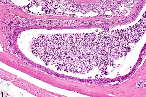
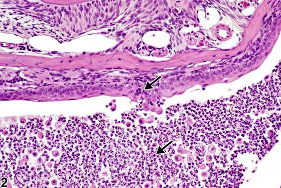
大鼠和小鼠的泪腺炎症可由多种原因引起,包括球后出血性外伤、感染性病原体(如大鼠的涎泪腺炎病毒)以及自身免疫性疾病。在NTP研究中,炎症有五个标准类别:急性、化脓性、慢性、慢性活动性和肉芽肿性。在急性炎症(Figure 1、Figure 2)中,主要浸润细胞是中性粒细胞,尽管也可能存在较少的巨噬细胞和淋巴细胞。还可能有水肿或充血的证据。中性粒细胞也是化脓性炎症中的主要浸润细胞类型,然而,在化脓性炎症中,中性粒细胞聚集,其中许多发生变性(化脓性渗出物)。渗出物中还可能存在细胞碎片(来自 resident 细胞群和浸润白细胞)、含纤维蛋白的蛋白性液体、较少的巨噬细胞、偶见的淋巴细胞或浆细胞,以及可能的感染性病原体。肉眼观察,这些病变将以脓液的存在为特征。在渗出物周围的组织中,可能存在成纤维细胞、纤维结缔组织和混合性炎症细胞,具体取决于病变的慢性程度。淋巴细胞在慢性炎症中占主导地位。淋巴细胞在慢性活动性炎症(Figure 3、Figure 4)中也占主导地位,但在慢性活动性炎症中,还存在大量中性粒细胞。两种病变均可能含有巨噬细胞。肉芽肿性炎症是慢性炎症的另一种形式,但该诊断要求存在大量聚集的、大的活化巨噬细胞、上皮样巨噬细胞或多核巨细胞。炎症与细胞浸润的区分在于是否存在其他变化,如水肿、出血、变性、坏死或其他组织损伤的证据。
诊断与分级建议(NTP 共识)
泪腺炎症只要存在就应诊断并赋予严重程度分级。应在诊断中通过适当的修饰词(急性、慢性等)指明炎症类型。应在病理描述中指明炎症在腺体内的位置(腺泡或导管)。相关病变(如纤维化)除非严重程度需要,否则不应单独诊断,但应在病理描述中描述。相关链接:泪腺 - 细胞浸润,单核细胞
图谱
英文原文
展开 NTP 官方英文描述与建议
Comment:
Lacrimal gland inflammation in rats and mice can result from various causes, including retrobulbar bleeding trauma, infectious agents such as sialodacryoadenitis virus in rats, and autoimmune disease. In NTP studies, there are five standard categories of inflammation: acute, suppurative, chronic, chronic-active, and granulomatous. In acute inflammation ( Figure 1 and Figure 2 ), the predominant infiltrating cell is the neutrophil, though fewer macrophages and lymphocytes may also be present. There may also be evidence of edema or hyperemia. The neutrophil is also the predominant infiltrating cell type in suppurative inflammation, however, in suppurative inflammation, the neutrophils are aggregated and many of them are degenerate (suppurative exudate). Cell debris, both from the resident cell populations and infiltrating leukocytes, proteinaceous fluid containing fibrin, fewer macrophages, occasional lymphocytes or plasma cells, and, possibly, an infectious agent may also be present in within the exudate. Grossly, these lesions would be characterized by the presence of pus. In the tissue surrounding the exudate, there may be fibroblasts, fibrous connective tissue, and mixed inflammatory cells, depending on the chronicity of the lesion. Lymphocytes predominate in chronic inflammation. Lymphocytes also predominate in chronic-active inflammation ( Figure 3 and Figure 4 ), but in chronic-active inflammation, there are also a significant number of neutrophils. Both lesions may contain macrophages. Granulomatous inflammation is another form of chronic inflammation, but this diagnosis requires the presence of a significant number of aggregated, large, activated macrophages, epithelioid macrophages, or multinucleated giant cells. Inflammation is differentiated from cellular infiltrates by the presence of other changes, such as edema, hemorrhage, degeneration, necrosis, or other evidence of tissue damage.
Recommendation:
Lacrimal gland inflammation should be diagnosed and assigned a severity grade whenever present. The type of inflammation should be indicated in the diagnosis with an appropriate modifier (acute, chronic, etc.). The location of the inflammation within the gland (acini or duct) should be indicated in the pathology narrative. Associated lesions (e.g., fibrosis) should not be diagnosed separately unless warranted by severity, but should be described in the pathology narrative. Related Links: Lacrimal Gland - Infiltration Cellular, Mononuclear Cell
来源与引用
原始条目:https://ntp.niehs.nih.gov/atlas/nnl/special-senses-system/lacrimal-gland/Inflammation
图谱总目录:NTP Nonneoplastic Lesion Atlas。 内容属美国联邦政府作品(公有领域),可引用;引用时请标注来源。
建议引用格式:NTP Nonneoplastic Lesion Atlas. Inflammation (lacrimal-gland). LabSmart 中文版:
https://www.labsmart.cn/ntp/lacrimal-gland-inflammation.html
中文图谱总页(可展开对照全部 595 条):https://www.labsmart.cn/ntp-atlas.html#lacrimal-gland-inflammation