血管扩张Angiectasis · 淋巴结 · 免疫系统 · NTP ntp-0014
诊断要点:只要淋巴结中出现血管扩张,就应作出诊断并赋予严重程度分级。
病变描述
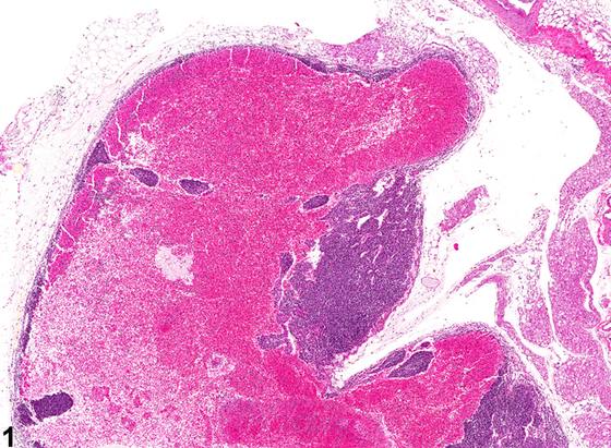
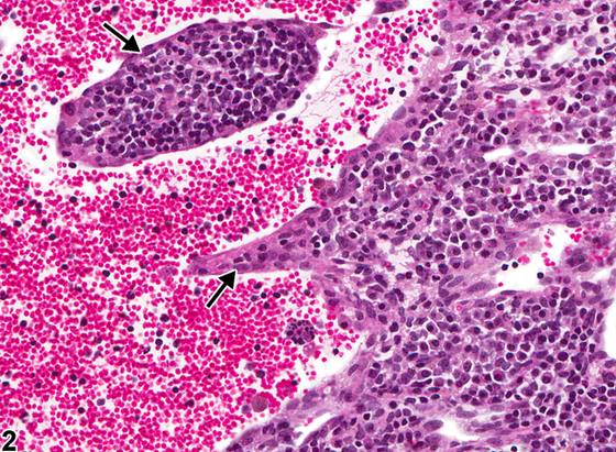
淋巴结血管扩张(angiectasis)的特征是皮质、髓质、被膜、门部或周围结缔组织内薄壁静脉的扩张(Figure 1)。血管腔由无明显异常的内皮细胞衬覆,通常充满血液。血管数量无明显增加。该病变最常见于某些小鼠和大鼠品系的肠系膜淋巴结,可能不伴有出血。血管扩张应与充血(congestion)、窦性红细胞增多(sinus erythrocytosis)、出血(hemorrhage)、血管瘤(hemangioma)和血管肉瘤(hemangiosarcoma)相鉴别。血管扩张与充血之间的区分有时较为困难,但充血时血管扩张的程度被认为是生理性正常的。血管扩张与出血不同;血管扩张时,血液局限于内皮衬覆的血管腔内(Figure 2,箭头),而出血时血液位于血管腔外。血管扩张可能与窦性红细胞增多难以区分,因为扩张的血管可类似于扩张并充满血液的淋巴管。应尝试区分血管扩张与血管瘤,尽管两者之间的差异并不总是显而易见。血管瘤往往为边界清楚、无包膜的肿块,由紧密排列的扩张血管腔构成。每个血管腔均为封闭的,由单层外观正常的内皮细胞衬覆,内皮细胞排列于胶原性间隔上,这些间隔通常较薄,但部分具有宽阔的胶原性间质。血管扩张通常不表现为边界清楚的肿块;扩张的血管通道常不规则地穿行于造血组织中。此外,血管扩张的扩张血管通道不具有侵袭性,也不显示内皮细胞异型性,借此可将该病变与血管肉瘤相区分。血管扩张以往的术语包括“hemangiectasis”“hemangiectasia”“vasodilation”和“vasodilatation”。
诊断与分级建议(NTP 共识)
只要淋巴结中出现血管扩张,就应作出诊断并赋予严重程度分级。
图谱
英文原文
展开 NTP 官方英文描述与建议
Comment:
Angiectasis of the lymph node is characterized by the dilatation of thin veins within the cortex, medulla, capsule, hilus, or surrounding connective tissue ( Figure 1 ). The vascular spaces are lined by unremarkable endothelial cells and are typically filled with blood. There is no apparent increase in the number of vessels. It is seen most often in the mesenteric lymph nodes of some mouse and rat strains and may not be associated with hemorrhage. Angiectasis should be distinguished from congestion, sinus erythrocytosis, hemorrhage, hemangioma, and hemangiosarcoma. The distinction between angiectasis and congestion is sometimes difficult, but with congestion, the degree of vascular dilation is considered physiologically normal. Angiectasis differs from hemorrhage; with angiectasis, the blood is contained within endothelium-lined vascular spaces ( Figure 2 , arrows), whereas with hemorrhage, the blood is outside the vascular spaces. Angiectasis may be difficult to differentiate from sinus erythrocytosis because dilated blood vessels can resemble dilated and blood-filled lymphatic vessels. The distinction between angiectasis and hemangioma should be attempted, although the difference is not always obvious. Hemangiomas tend to be well-circumscribed, unencapsulated masses composed of tightly packed, dilated vascular spaces. Each vascular space is enclosed and lined by a single layer of normal-appearing endothelial cells aligned on collagenous septa, which are usually thin, although some have broad collagenous stroma. Angiectasis does not usually present as a well-circumscribed mass; the dilated vascular channels often course irregularly through the hematopoietic tissue. Furthermore, the dilated vascular channels of angiectasis are not invasive and do not demonstrate endothelial cell atypia, which differentiates this lesion from hemangiosarcoma. Previous terms for angiectasis include "hemangiectasis," "hemangiectasia," "vasodilation," and "vasodilatation."
Recommendation:
Whenever angiectasis is present in lymph nodes, it should be diagnosed and assigned a severity grade.
来源与引用
原始条目:https://ntp.niehs.nih.gov/atlas/nnl/immune-system/lymph-node/Angiectasis
图谱总目录:NTP Nonneoplastic Lesion Atlas。 内容属美国联邦政府作品(公有领域),可引用;引用时请标注来源。
建议引用格式:NTP Nonneoplastic Lesion Atlas. Angiectasis (lymph-node). LabSmart 中文版:
https://www.labsmart.cn/ntp/lymph-node-angiectasis.html
中文图谱总页(可展开对照全部 595 条):https://www.labsmart.cn/ntp-atlas.html#lymph-node-angiectasis