组织细胞性细胞浸润Inflitration Cellular Histiocyte · 淋巴结 · 免疫系统 · NTP ntp-0010
诊断要点:大鼠和小鼠淋巴结中组织细胞增多无论何时出现,均应作出诊断并给出严重程度分级。该病变应始终结合受试物、处理组、给药途径和受累淋巴结(与同期对照比较)进行诊断。
病变描述
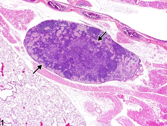
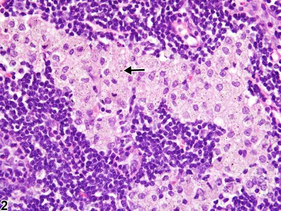
大鼠和小鼠淋巴结中的组织细胞(histiocyte)可能增多。该病变可自发发生(例如,肠系膜淋巴结受到日粮抗原刺激),也可继发于受试物处理。组织细胞增多可能反映驻留窦组织细胞的增殖,或经输入淋巴管进入的组织细胞增多。该病变可位于被膜下窦、副皮质区窦或髓窦内(Figure 1,箭头)。若该病变在特定剂量组的同一淋巴结(例如吸入研究中的支气管淋巴结)中发生,则可能提示与处理相关的效应。受累淋巴结中的组织细胞胞质内可能含有不溶性颗粒物质,如被吞噬的受试物或赋形剂物质(Figure 2,箭头)或色素(含铁血黄素等)。在持续自发抗原或异物刺激下的淋巴结(例如肠系膜淋巴结)中,组织细胞增多必须与处理所致的增多相鉴别。淋巴结组织细胞既往使用的术语为“组织细胞增生(histiocyte hyperplasia)”和“组织细胞浸润(histiocyte infiltrate)”。
诊断与分级建议(NTP 共识)
大鼠和小鼠淋巴结中组织细胞增多无论何时出现,均应作出诊断并给出严重程度分级。该病变应始终结合受试物、处理组、给药途径和受累淋巴结(与同期对照比较)进行诊断。
图谱
英文原文
展开 NTP 官方英文描述与建议
Comment:
Histiocytes may be increased in the lymph nodes of rats and mice. This lesion can occur spontaneously (e.g., dietary antigenic stimulation of mesenteric lymph nodes) or secondary to treatment. Increases may reflect proliferation of resident sinusoidal histiocytes or an increased influx of histiocytes via the afferent lymphatics. This lesion may be within subcapsular, paracortical, or medullary sinuses ( Figure 1 , arrows). If this lesion occurs in the same lymph node for a particular dose group (e.g., bronchial lymph node in an inhalation study), it may suggest a treatment-related effect. Histiocytes in affected lymph nodes may contain intracytoplasmic insoluble particulate matter, such as phagocytized test article or vehicle material ( Figure 2 , arrow) or pigment (hemosiderin, etc.). Histiocyte increases in lymph nodes under constant spontaneous antigenic or foreign material stimulation (e.g., mesenteric lymph nodes) must be differentiated from increases due to treatment. Previous terms for histiocytes in lymph nodes are "histiocyte hyperplasia" and "histiocyte infiltrate."
Recommendation:
Whenever present, histiocyte increases in the lymph nodes of rats and mice should be diagnosed and given a severity grade. This lesion should always be diagnosed in the context of the test article, treatment group, route of administration, and lymph node involved (compare with concurrent controls).
来源与引用
原始条目:https://ntp.niehs.nih.gov/atlas/nnl/immune-system/lymph-node/InflitrationCellularHistiocyte
图谱总目录:NTP Nonneoplastic Lesion Atlas。 内容属美国联邦政府作品(公有领域),可引用;引用时请标注来源。
建议引用格式:NTP Nonneoplastic Lesion Atlas. Inflitration Cellular Histiocyte (lymph-node). LabSmart 中文版:
https://www.labsmart.cn/ntp/lymph-node-inflitration-cellular-histiocyte.html
中文图谱总页(可展开对照全部 595 条):https://www.labsmart.cn/ntp-atlas.html#lymph-node-inflitration-cellular-histiocyte