角化性囊性增生Hyperplasia Cystic Keratinizing · 口腔黏膜 · 消化系统 · NTP ntp-0226
诊断要点:无论何时出现,囊性角化性增生均应予以诊断,并根据充满角蛋白的囊样区域的数量和大小进行分级。
病变描述
囊性角化性增生(cystic keratinizing hyperplasia)的特征是牙龈区域上皮显著增生,并呈乳头状皱褶隆起,在牙龈及邻近组织中形成充满角蛋白的囊样腔隙。部分此类病变为孤立性发生,而另一些则与黏膜的弥漫性(diffuse)增生病灶相伴发生。腔隙的数量可从单个至多个不等,使病变呈现多房外观。通常可见囊性结构与上覆的牙龈口腔黏膜之间存在连接,提示囊性角化性增生可能起源于鳞状上皮增生。该病变的鉴别诊断包括鳞状囊肿(squamous cyst,见口腔黏膜-囊肿,鳞状)和牙周袋(periodontal pocket,见牙齿-牙周袋)。鳞状囊肿和牙周袋通常为单房结构,衬以相当薄的、外观良性的鳞状上皮,且似乎并非起源于增生的牙龈黏膜。鳞状囊肿的囊壁鳞状上皮中可能存在毛囊。牙周袋是啮齿类动物常见的背景性改变,局限于牙齿周围的牙周组织,尤其是磨牙。牙周袋代表异常加深的牙龈沟,衬以角化的鳞状上皮,其腔内可能含有毛发、饲料、垫料和角蛋白。囊性角化性增生还必须与早期鳞状细胞癌相鉴别,后者的细胞呈多形性或非典型性,且鳞状细胞成熟紊乱。囊性角化性增生可能处于增生与鳞状细胞癌的连续谱系上,因为在一项研究中观察到该病变,同时该研究中牙龈鳞状细胞癌也显著增加。
诊断与分级建议(NTP 共识)
无论何时出现,囊性角化性增生均应予以诊断,并根据充满角蛋白的囊样区域的数量和大小进行分级。继发性病变,如炎症或骨重塑,除非是病变的重要组成部分,否则不应单独诊断。相关链接:口腔黏膜-囊肿,鳞状;牙齿-牙周袋
图谱
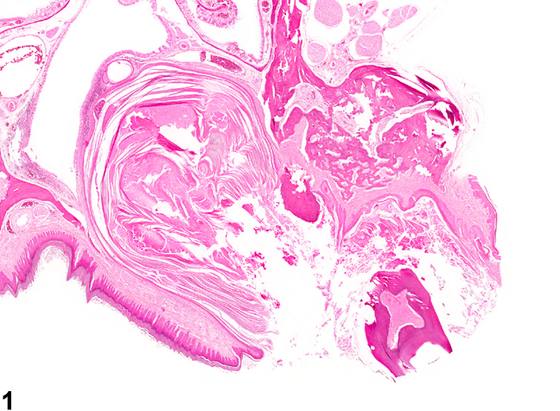
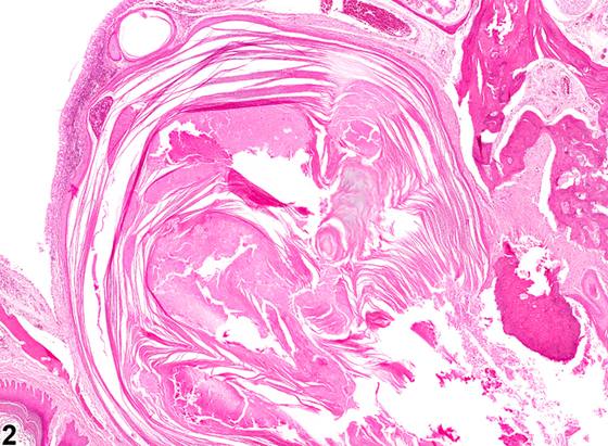
英文原文
展开 NTP 官方英文描述与建议
Comment:
Cystic keratinizing hyperplasia is characterized by areas in the gingiva in which the epithelium is markedly hyperplastic and raised in papillary folds forming cyst-like spaces filled with keratin in the gingival and adjacent tissues. Some of these lesions occur in isolation, whereas others occur in association with diffuse hyperplastic lesions of the mucosa. The number of cavities can vary from one to multiple, giving the lesion a multilocular appearance. Generally, a connection can be seen between the cystic structure and the overlying gingival oral mucosa, suggesting the cystic keratinizing hyperplasia may have arisen from squamous hyperplasia. A differential diagnosis for this lesion is a squamous cyst (see Oral Mucosa - Cyst, Squamous ) and periodontal pocket (see Tooth - Periodontal Pocket ). Squamous cysts and periodontal pockets tend to be unilocular structures lined by a fairly thin, benign-appearing squamous epithelium and do not appear to be arising from a hyperplastic gingival mucosa. Squamous cysts may have hair follicles in the squamous epithelium lining the cyst. Periodontal pockets are common background alterations in rodents restricted to the periodontal tissues surrounding the teeth, especially molars. The pockets represent an abnormally deepened gingival sulcus that is lined by keratinized squamous epithelium, and the lumen may contain hair, feed, bedding material, and keratin. Cystic keratinizing hyperplasia must also be differentiated from early squamous cell carcinoma, in which the cells are pleomorphic or atypical and there is disorderly maturation of the squamous cells. Cystic keratinizing hyperplasia may be on a continuum with hyperplasia and squamous cell carcinoma as this lesion was seen in a study in which there was also a significant increase in gingival squamous cell carcinomas.
Recommendation:
Whenever present, cystic keratinizing hyperplasia should be diagnosed and graded based on the number and size of cyst-like areas filled with keratin. Secondary lesions, such as inflammation or bone remodeling, should not be diagnosed separately unless they are a significant component of the lesion. Related Links: Oral Mucosa - Cyst, Squamous Tooth - Periodontal Pocket
来源与引用
原始条目:https://ntp.niehs.nih.gov/atlas/nnl/alimentary-system/oral-mucosa/HyperplasiaCysticKeratinizing
图谱总目录:NTP Nonneoplastic Lesion Atlas。 内容属美国联邦政府作品(公有领域),可引用;引用时请标注来源。
建议引用格式:NTP Nonneoplastic Lesion Atlas. Hyperplasia Cystic Keratinizing (oral-mucosa). LabSmart 中文版:
https://www.labsmart.cn/ntp/oral-mucosa-hyperplasia-cystic-keratinizing.html
中文图谱总页(可展开对照全部 595 条):https://www.labsmart.cn/ntp-atlas.html#oral-mucosa-hyperplasia-cystic-keratinizing