合胞体巨细胞Syncytial Giant Cell · 甲状旁腺 · 内分泌系统 · NTP ntp-0053
诊断要点:推荐对甲状旁腺合体巨细胞形成作出诊断。不需要进行严重程度分级。若两侧甲状旁腺均受累,诊断应限定为双侧性。
病变描述
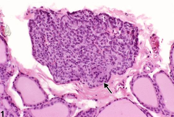
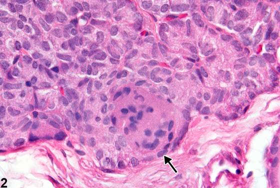
大鼠甲状旁腺中可出现多核合体细胞(multinucleated syncytial cells)。其数量不一,但这些巨细胞可占据腺体达一半。它们由相邻主细胞(chief cells)的胞质融合形成。胞质可呈致密嗜酸性。合体巨细胞被认为不干扰甲状旁腺功能。对于这是否为生前变化尚无共识。一些科学家认为这是死后人为假象。
诊断与分级建议(NTP 共识)
推荐对甲状旁腺合体巨细胞形成作出诊断。不需要进行严重程度分级。若两侧甲状旁腺均受累,诊断应限定为双侧性。
图谱
英文原文
展开 NTP 官方英文描述与建议
Comment:
Multinucleated syncytial cells occur in the parathyroid of rats. Their number varies, but these giant cells may occupy up to one-half of the gland. They are formed by cytoplasmic fusion of adjacent chief cells. The cytoplasm may be densely eosinophilic. Syncytial giant cells are not believed to interfere with parathyroid function. There is no consensus that this is an antemortem change. Some scientists consider this a postmortem artifact.
Recommendation:
Diagnosis of syncytial giant cell formation in the parathyroid is recommended. A severity grade is not needed. If both parathyroids are involved, the diagnosis should be qualified as bilateral.
来源与引用
原始条目:https://ntp.niehs.nih.gov/atlas/nnl/endocrine-system/parathyroid-gland/SyncytialGiantCell
图谱总目录:NTP Nonneoplastic Lesion Atlas。 内容属美国联邦政府作品(公有领域),可引用;引用时请标注来源。
建议引用格式:NTP Nonneoplastic Lesion Atlas. Syncytial Giant Cell (parathyroid-gland). LabSmart 中文版:
https://www.labsmart.cn/ntp/parathyroid-gland-syncytial-giant-cell.html
中文图谱总页(可展开对照全部 595 条):https://www.labsmart.cn/ntp-atlas.html#parathyroid-gland-syncytial-giant-cell