上皮变性Epithelium Degeneration · 前列腺 · 雄性生殖系统 · NTP ntp-0442
诊断要点:存在时,该病变应当诊断并分级。化学物质所致的加重值得在病理学叙述中记录。如有可能,应确定受累的叶,并在组织标识中指明。
病变描述
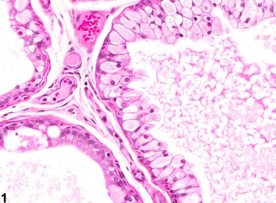
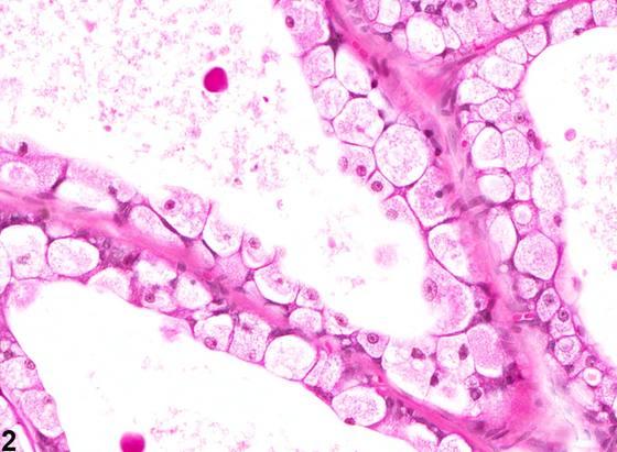
上皮变性(epithelial degeneration)的特征是增大的腺泡上皮细胞胞质呈颗粒状至泡沫状改变,这些细胞形成单层衬覆层(Figure 1、Figure 2),或脱落并阻塞腺泡腔(Figure 3)。受累细胞增大,胞质富含细颗粒状物质,可发生破裂和解体。上皮变性可为局灶性(focal)或多灶性(multifocal)。该病变可见于任何叶,但最常见于腹侧叶。上皮变性在老龄前列腺中是相当常见的观察所见。该病变可伴有炎性浸润。
诊断与分级建议(NTP 共识)
存在时,该病变应当诊断并分级。化学物质所致的加重值得在病理学叙述中记录。如有可能,应确定受累的叶,并在组织标识中指明。若成对的叶均受累,诊断应指明该变化为双侧性,严重程度依据受累更严重的叶判定。
图谱
英文原文
展开 NTP 官方英文描述与建议
Comment:
Epithelial degeneration is characterized by granular to foamy cytoplasm alteration of enlarged acinar epithelial cells that form a single lining layer ( Figure 1 and Figure 2 ) or exfoliate and occlude the lumen of acini ( Figure 3 ). Affected cells are enlarged with abundant finely granular cytoplasm and may rupture and disintegrate. Epithelial degeneration can be focal or multifocal. The lesion can be observed in any lobe, but is most commonly found in the ventral lobe. Epithelial degeneration is a fairly common observation in the aging prostate. The lesion may be accompanied by inflammatory infiltrates.
Recommendation:
When present, this lesion should be diagnosed and graded. Exacerbation by chemical agents warrants documentation in the pathology narrative. The affected lobe(s) should be identified if possible and indicated in the tissue identification. If paired lobes are involved, the diagnosis should indicate that the change is bilateral, with severity based on the more severely affected lobe.
来源与引用
原始条目:https://ntp.niehs.nih.gov/atlas/nnl/reproductive-system-male/prostate/Epithelium-Degeneration
图谱总目录:NTP Nonneoplastic Lesion Atlas。 内容属美国联邦政府作品(公有领域),可引用;引用时请标注来源。
建议引用格式:NTP Nonneoplastic Lesion Atlas. Epithelium Degeneration (prostate). LabSmart 中文版:
https://www.labsmart.cn/ntp/prostate-epithelium-degeneration.html
中文图谱总页(可展开对照全部 595 条):https://www.labsmart.cn/ntp-atlas.html#prostate-epithelium-degeneration