上皮增生Epithelium Hyperplasia · 前列腺 · 雄性生殖系统 · NTP ntp-0443
诊断要点:应记录增生的分布以及该病变的严重程度。如有可能,应确定受累的叶并在组织识别中注明(例如,prostate, ventral lobe, epithelium hyperplasia, 轻微(mild))。
病变描述
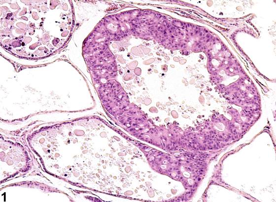
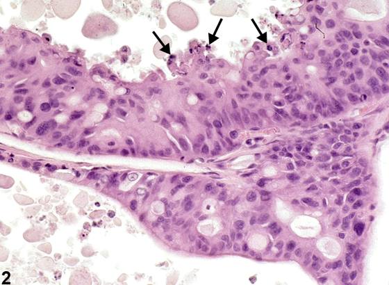
增生(hyperplasia)的特征是上皮腺泡衬覆细胞数量增多,通常突入腺泡腔。增生通常累及1至3个相邻腺泡,衬覆细胞可几乎完全充满腺泡腔,但不压迫相邻腺泡。增生的细胞呈立方状至高柱状,在大鼠通常具有颗粒状嗜酸性胞质。当存在多个孤立的分泌区域时,可呈多房外观(Figure 3、Figure 4)。随着增生细胞堆积,部分细胞发生变性、坏死或凋亡(箭头,Figure 2)。在大鼠皮下给予α-雄激素激动剂去氧肾上腺素(phenylephrine)可诱导前列腺增生,其特征为上皮细胞堆积,呈乳头状和筛状结构并出芽。已有报道,老年 brown Norway 大鼠前列腺背叶和腹叶出现与增生一致的自发性年龄依赖性细胞过度生长。前列腺增生见于老龄动物,可累及任何叶。与前列腺腺瘤的鉴别主要基于:腺瘤为膨胀性、相对离散的上皮增生充满腺泡并压迫相邻腺泡。相比之下,TRAMP 小鼠中所见的前列腺增生由深嗜碱性细胞堆积构成(箭头,Figure 5)。在 TRAMP 小鼠中,部分研究者将这些病变视为前列腺上皮内瘤变(prostatic intraepithelial neoplasia)病变。
诊断与分级建议(NTP 共识)
应记录增生的分布以及该病变的严重程度。如有可能,应确定受累的叶并在组织识别中注明(例如,prostate, ventral lobe, epithelium hyperplasia, 轻微(mild))。如果成对的叶均受累,诊断应限定为双侧性,严重程度评分以受累更严重的叶确定。任何可能治疗相关的增生增加均应记录在病理学叙述中,并与其他生殖和内分泌组织的相关变化相联系。
图谱
英文原文
展开 NTP 官方英文描述与建议
Comment:
Hyperplasia is characterized by an increased number of epithelial acinar lining cells that typically project into the acinar lumen. Hyperplasia typically affects one to three contiguous acini, and the lining cells may almost completely fill the acinar lumen but do not compress contiguous acini. The hyperplastic cells range from cuboidal to tall columnar and in the rat usually have a granular eosinophilic cytoplasm. There may be a multilocular appearance when there are multiple isolated areas of secretion ( Figure 3 and Figure 4 ). As the hyperplastic cells pile up, some undergo degeneration and necrosis or apoptosis (arrow, Figure 2 ). Subcutaneous administration of the α-androgenic agonist phenylephrine in rats induces prostatic hyperplasia characterized by piling up of epithelial cells, with papillary and cribriform patterns and budding out. Spontaneous age-dependent overgrowth of cells, consistent with hyperplasia, in the dorsal and ventral lobes of prostate in old brown Norway rats has been reported. Prostatic hyperplasia is seen in aging animals and can involve any lobe. Distinction from prostatic adenoma is based largely on compression of contiguous acini by an expanding and relatively discrete epithelial proliferation filling acini. By way of contrast, the prostatic hyperplasia seen in the TRAMP mouse consists of piling up of hyperbasophilic cells (arrows, Figure 5 ). In the TRAMP mouse these are considered prostatic intraepithelial neoplasia lesions by some investigators.
Recommendation:
The distribution of hyperplasia should be recorded, along with the severity of this lesion. The affected lobe(s) should be identified if possible and indicated in the tissue identification (e.g., prostate, ventral lobe, epithelium hyperplasia, mild). If paired lobes are affected, the diagnosis should be qualified as bilateral and the severity score determined by the more severely affected lobe. Any potentially treatment-related increase in hyperplasia should be documented in the pathology narrative and correlated with related changes in other reproductive and endocrine tissues.
来源与引用
原始条目:https://ntp.niehs.nih.gov/atlas/nnl/reproductive-system-male/prostate/Epithelium-Hyperplasia
图谱总目录:NTP Nonneoplastic Lesion Atlas。 内容属美国联邦政府作品(公有领域),可引用;引用时请标注来源。
建议引用格式:NTP Nonneoplastic Lesion Atlas. Epithelium Hyperplasia (prostate). LabSmart 中文版:
https://www.labsmart.cn/ntp/prostate-epithelium-hyperplasia.html
中文图谱总页(可展开对照全部 595 条):https://www.labsmart.cn/ntp-atlas.html#prostate-epithelium-hyperplasia