萎缩Atrophy · 骨骼肌 · 骨骼肌肉系统 · NTP ntp-0594
诊断要点:当存在时,骨骼肌萎缩应当被诊断并分级。未受累纤维的代偿性肥大无需单独记录,除非其严重程度需要如此。
病变描述
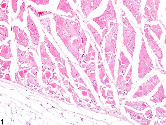
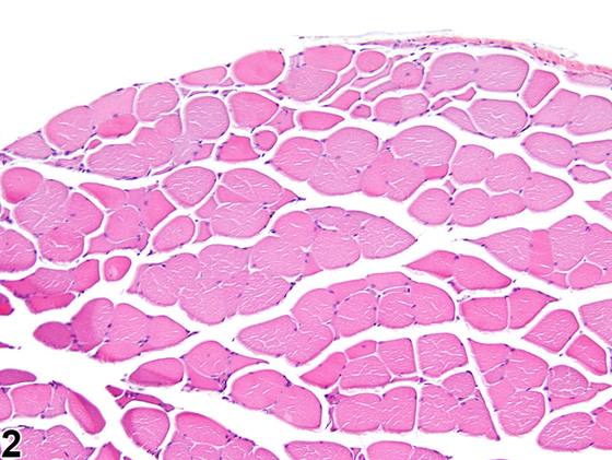
骨骼肌萎缩可由起源于骨骼肌纤维内部的退行性过程引起,也可继发于去神经支配(去神经性萎缩),或在老龄啮齿类动物中自发发生。它是任何导致肌纤维细胞器降解和/或丢失的损伤的常见反应。示例包括废用、恶病质、营养或代谢紊乱、血管功能不全、激素生长控制机制紊乱以及给予肌毒性外源化学物。无论病因如何,常同时存在存活/未受累肌纤维的代偿性肥大。骨骼肌萎缩的主要组织学特征是肌纤维直径减小(Figure 1、Figure 2)。受累肌纤维常呈圆形至棱角状,肌浆嗜酸性增强。在去神经性萎缩中,特征性组织学表现为受压的棱角状肌纤维伴细胞核密集。非神经源性原因所致的萎缩,例如由给予肌毒性外源化学物所诱导者,通常缺乏这些纤维,而是伴有传统肌病性改变,如坏死性或玻璃样变肌纤维、撕裂或碎裂的肌纤维以及具有中央核的肌纤维。老龄啮齿类动物的自发性萎缩(常称为后肢肌病)的组织学特征为肌纤维大小和数量减少;肌纤维大小变异增加;退行性包涵体、脂褐素和脂滴蓄积增加;以及结缔组织增加。常存在棱角状肌纤维,提示脊髓退行性变可能在该过程中发挥作用。然而,并非在所有受累动物中均一致观察到脊神经病变;其发病机制仍不清楚。该病变主要见于受累大鼠的后肢。已在包括Wistar和Sprague-Dawley在内的多个大鼠品系中描述,但尚未在F344大鼠中记录。萎缩可均匀累及肌纤维,或选择性靶向特定肌纤维类型。当萎缩与废用、恶病质或营养不良相关时,II型纤维被选择性累及。在大鼠中长期给予皮质类固醇会导致II型快肌纤维的选择性萎缩。相反,在实验性诱导的甲状腺毒症中,I型纤维被选择性靶向。
诊断与分级建议(NTP 共识)
当存在时,骨骼肌萎缩应当被诊断并分级。未受累纤维的代偿性肥大无需单独记录,除非其严重程度需要如此。萎缩的可能原因(例如原发性变性、继发于去神经支配,或老龄动物中的自发性)应在病理学叙述中讨论。受累肌纤维的组织学表现,特别是关于是否存在棱角状肌纤维,应纳入叙述中。当其严重程度需要时,其他肌病性改变,如肌纤维坏死或变性,应当单独记录并分级。
图谱
英文原文
展开 NTP 官方英文描述与建议
Comment:
Skeletal muscle atrophy can occur due to degenerative processes originating within the skeletal muscle fibers, secondary to denervation (denervation atrophy), or spontaneously in aging rodents. It is a common reaction to any injury that results in the degradation and/or loss of myofiber organelles. Examples include disuse, cachexia, nutritional or metabolic derangements, vascular insufficiency, disturbances of hormonal growth control mechanisms, and administration of myotoxic xenobiotics. Compensatory hypertrophy of surviving/unaffected myofibers is often present concomitantly, regardless of the cause. The main histologic feature of skeletal muscle atrophy is a reduction in myofiber diameter ( Figure 1 and Figure 2 ). Affected myofibers are often rounded to angular with hypereosinophilic sarcoplasm. In denervation atrophy, the characteristic histologic feature is that of compressed angular myofibers with crowded nuclei. Atrophy due to nonneurogenic causes, such as that induced by the administration of myotoxic xenobiotics, often lacks these fibers and is instead accompanied by traditional myopathic changes, such as necrotic or hyalinized myofibers, split or fragmented myofibers, and myofibers with central nuclei. Spontaneous atrophy of aging rodents (often referred to as hind leg myopathy) is histologically characterized by decreased myofiber size and number; increased myofiber size variation; increased accumulation of degenerative inclusion bodies, lipofuscin, and lipid droplets; and increased connective tissue. Angulated myofibers are often present, suggesting a possible role of spinal degeneration within this process. However, spinal nerve lesions are not consistently observed in all affected animals; the pathogenesis remains unclear. This condition is predominately noted in the hind limbs of affected rats. It has been described in several strains of rats, including Wistar and Sprague-Dawley, but has not been recorded in the F344 rat. Atrophy can uniformly affect myofibers or selectively target specific muscle fiber types. Type II fibers are selectively affected when atrophy is associated with disuse, cachexia, or malnutrition. Chronic administration of corticosteroids in rats results in the selective atrophy of type II fast-twitch fibers. Conversely, type I fibers are selectively targeted during experimentally induced thyrotoxicosis.
Recommendation:
When present, skeletal muscle atrophy should be diagnosed and graded. Compensatory hypertrophy of unaffected fibers does not need to be recorded separately unless warranted by severity. Potential causes of the atrophy (e.g., primary degeneration, secondary to denervation, or spontaneously in aging animals) should be discussed in the pathology narrative. The histologic appearance of the affected myofibers, specifically regarding the presence of angulated myofibers, should be included in the narrative. When warranted by severity, additional myopathic changes, such as myofiber necrosis or degeneration, should be recorded and graded separately.
来源与引用
原始条目:https://ntp.niehs.nih.gov/atlas/nnl/musculoskeletal-system/skeletal-muscle/Atrophy
图谱总目录:NTP Nonneoplastic Lesion Atlas。 内容属美国联邦政府作品(公有领域),可引用;引用时请标注来源。
建议引用格式:NTP Nonneoplastic Lesion Atlas. Atrophy (skeletal-muscle). LabSmart 中文版:
https://www.labsmart.cn/ntp/skeletal-muscle-atrophy.html
中文图谱总页(可展开对照全部 595 条):https://www.labsmart.cn/ntp-atlas.html#skeletal-muscle-atrophy