变性Degeneration · 骨骼肌 · 骨骼肌肉系统 · NTP ntp-0586
诊断要点:当存在时,肌纤维变性应予以诊断并分级。如果一项研究中同时存在变性和坏死,应诊断占优势的病变,并在病理学叙述中描述另一病变。
病变描述
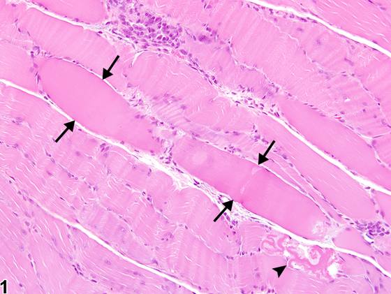
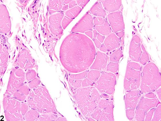
变性的肌肉在大体上可表现为苍白或暗色。在组织学上,变性的肌纤维可表现出多种显微镜下变化,包括细胞肿胀、嗜酸性增强(hypereosinophilia)、空泡化、横纹消失、碎裂和纤维断裂(Figure 1、Figure 2、Figure 3、Figure 4、Figure 5、Figure 6、Figure 7)。断裂的纤维通常可通过“回缩帽”(retraction caps)的存在来识别,回缩帽是断裂纤维游离端的凹陷。巨噬细胞常可见与变性肌纤维密切相伴,因为它们在吞噬相关碎片中发挥重要作用(Figure 4)。由于肌纤维较长,变性通常仅累及纤维的一个或数个节段,而非整个肌纤维。变性区域可不同程度地伴有其他肌病性改变,如坏死、萎缩、代偿性肥大、再生和纤维化(Figure 5)。无论病因如何,变性都是肌纤维损伤的常见后果。常见病因包括化学刺激物/肌毒素、代谢异常、创伤和感染。与其他组织一样,变性可以是可逆的;然而,如果损伤性刺激持续存在,将达到“不可逆点”(point of no return)。当这种情况发生时,变性过程变为不可逆,随后出现肌纤维坏死。变性和坏死代表一个连续的病变谱系,因此在一个给定病变中二者常同时存在。由于骨骼肌反应方式有限,它们具有相似的形态学特征,因此在组织学上可能难以区分。这会造成诊断上的困难。在评估毒性研究时,病理学家必须为这两种病变建立明确的判定标准,并在应用时保持一致和审慎。标准应在叙述中加以描述。“肌病”(myopathy)一词通常用于描述以变性和坏死为关键特征的骨骼肌疾病。然而,由于肌病是一个通用术语,且通常涵盖一组病变而非某一独立病变,因此不建议使用该词。
诊断与分级建议(NTP 共识)
当存在时,肌纤维变性应予以诊断并分级。如果一项研究中同时存在变性和坏死,应诊断占优势的病变,并在病理学叙述中描述另一病变。然而,如果同时记录两种病变最能描述所发生的变化,病理学家可以记录两者。伴随的再生应单独记录,当其存在达到显著程度时赋予严重程度分级。其他继发性病变,如炎症和出血,除非严重程度确有必要,否则不应单独诊断,但应在叙述中描述。相关链接:骨骼肌 - 坏死
图谱
英文原文
展开 NTP 官方英文描述与建议
Comment:
Degenerated muscle can grossly appear either pale or dark. Histologically, degenerating myofibers can exhibit a variety of microscopic changes, including cell swelling, hypereosinophilia, vacuolation, loss of striation, fragmentation, and rupture of fibers ( Figure 1 , Figure 2 , Figure 3 , Figure 4 , Figure 5 , Figure 6 , and Figure 7 ). Ruptured fibers can often be identified by the presence of "retraction caps," which are concavities at the free ends of the ruptured fibers. Macrophages are often noted in close association with degenerating myofibers, as they play an important role in phagocytizing associated debris ( Figure 4 ). Due to the long length of muscle fibers, degeneration often only affects a segment or several segments of the fiber, as opposed to the entire myofiber. Regions of degeneration can be variably accompanied by additional myopathic changes, such as necrosis, atrophy, compensatory hypertrophy, regeneration, and fibrosis ( Figure 5 ). Degeneration is a common sequela of myofiber injury, regardless of the cause. Common causes include chemical irritants/myotoxins, abnormal metabolism, trauma, and infection. As in other tissues, degeneration can be reversible; however, if the injurious stimulus persists, a "point of no return" will be reached. When this occurs, the degenerative process becomes irreversible and myofiber necrosis follows. Degeneration and necrosis represent a continuum of lesions and therefore are often both present within a given lesion. Due to the limited repertoire of skeletal muscle responses, they share similar morphologic features and can thus be difficult to differentiate histologically. This can create a diagnostic challenge. When evaluating a toxicity study, it is important for the pathologist to establish distinct criteria for both lesions and to be consistent and careful when applying them. Criteria should be described in the narrative. The term "myopathy" is commonly used to describe disorders of skeletal muscle in which degeneration and necrosis are key features. However, since myopathy is a general term and one that typically encompasses a collection of lesions rather than one distinct lesion, its use is not recommended.
Recommendation:
When present, myofiber degeneration should be diagnosed and graded. If both degeneration and necrosis are present in a study, the predominant lesion should be diagnosed and the other described in the pathology narrative. However, the pathologist may record both lesions if it best describes the changes that are occurring. Concomitant regeneration should be recorded separately and assigned a severity grade when present to a significant extent. Other secondary lesions, such as inflammation and hemorrhage, should not be diagnosed separately unless warranted by severity but should be described in the narrative. Related Links: Skeletal Muscle - Necrosis
来源与引用
原始条目:https://ntp.niehs.nih.gov/atlas/nnl/musculoskeletal-system/skeletal-muscle/Degeneration
图谱总目录:NTP Nonneoplastic Lesion Atlas。 内容属美国联邦政府作品(公有领域),可引用;引用时请标注来源。
建议引用格式:NTP Nonneoplastic Lesion Atlas. Degeneration (skeletal-muscle). LabSmart 中文版:
https://www.labsmart.cn/ntp/skeletal-muscle-degeneration.html
中文图谱总页(可展开对照全部 595 条):https://www.labsmart.cn/ntp-atlas.html#skeletal-muscle-degeneration