背根神经节神经元空泡化Dorsal Root Ganglion Neuron Vacuolation · 脊髓 · 神经系统 · NTP ntp-0380
诊断要点:当出现时,神经元空泡化在NTP研究中应予诊断,记录其亚部位,并进行严重程度分级。
病变描述
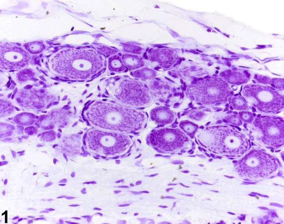
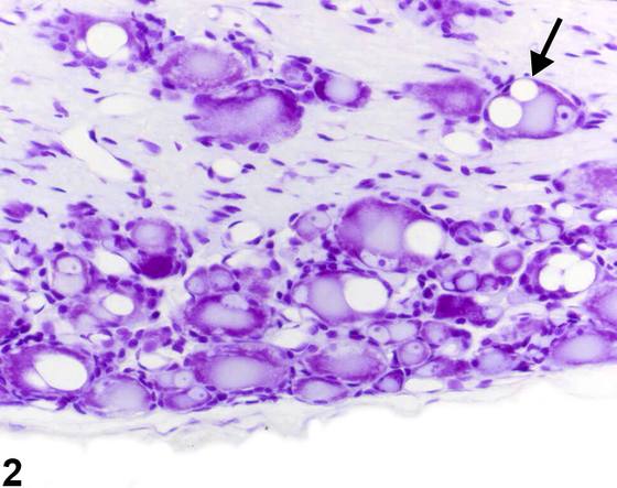
神经元空泡化(neuronal vacuolation)虽可见于任何发生变性的神经元群体,但其在背根神经节(dorsal root ganglia)中的神经病理学意义尤受关注。Figure 1显示了大鼠背根神经节神经元的正常形态。诊断上的问题在于要认识到,这些神经节部位的组织未接触毒性化合物时,神经元空泡化偶尔也可以低发生率出现,并且在老年啮齿类动物中更为常见。重要的是,神经元空泡化可能是毒性的显著效应。空泡表现为受累神经元核周体(perikaryon)内大小不一但通常较大的透明空泡,可伴有染色质溶解(chromatolysis)(Figure 2,箭头)。空泡的衬里由单层膜构成,该膜有短突起伸入空泡内,并伴有染色质溶解。电子显微镜图像未能明确识别受累的肿胀细胞器,但滑面内质网和高尔基体是最可能的候选者。鉴于已知神经元空泡化可作为偶发性 incidental finding 出现,仔细检查对照和治疗组在相应区域是否存在该变化十分重要。相关变化的存在,如神经元染色质溶解或胶质细胞或卫星细胞的邻近反应,可能有助于将空泡化归因为一种病变。
诊断与分级建议(NTP 共识)
当出现时,神经元空泡化在NTP研究中应予诊断,记录其亚部位,并进行严重程度分级。存在并发病变时,通常诊断严重程度最高的病变。其他并发病变,若严重程度足以支持,可单独诊断。
图谱
英文原文
展开 NTP 官方英文描述与建议
Comment:
Neuronal vacuolation, while it may be seen in any population of neurons undergoing degeneration, is a particular concern for its neuropathologic significance in the dorsal root ganglia. Figure 1 shows the normal appearance of rat dorsal root ganglionic neurons. At issue diagnostically is the recognition that neuronal vacuolation at these ganglionic sites may occasionally be present at a low incidence in tissue not exposed to toxic compounds and is more common in older rodents. Importantly, neuronal vacuolation may be a significant effect of toxicity. The vacuoles appear as variably sized, but generally large clear vacuoles in the perikaryon of the affected neurons may be accompanied by chromatolysis ( Figure 2 , arrow). The lining of the vacuoles is composed of a single membrane, and short protrusions of this membrane occur into the vacuoles, accompanied by chromatolysis. Electron microscopic images do not clearly identify the affected swollen organelles, but smooth endoplasmic reticulum and Golgi apparatus are the most likely candidates. In view of the known occurrence of neuronal vacuolation as an occasional incidental finding, it is important to carefully examine control and treatment groups for its presence in comparable regions. The existence of associated changes such as neuronal chromatolysis or adjacent responses by glia or satellite cells may be helpful in attributing vacuolization as a lesion.
Recommendation:
When present, neuronal vacuolation is diagnosed in NTP studies, the subsite recorded, and the severity graded. In the presence of concurrent lesions, lesions with the most severity are typically diagnosed. Other concurrent lesions may be diagnosed separately, if warranted by the severity.
来源与引用
原始条目:https://ntp.niehs.nih.gov/atlas/nnl/nervous-system/spinal-cord/DorsalRootGanglionNeuron-Vacuolation
图谱总目录:NTP Nonneoplastic Lesion Atlas。 内容属美国联邦政府作品(公有领域),可引用;引用时请标注来源。
建议引用格式:NTP Nonneoplastic Lesion Atlas. Dorsal Root Ganglion Neuron Vacuolation (spinal-cord). LabSmart 中文版:
https://www.labsmart.cn/ntp/spinal-cord-dorsal-root-ganglion-neuron-vacuolation.html
中文图谱总页(可展开对照全部 595 条):https://www.labsmart.cn/ntp-atlas.html#spinal-cord-dorsal-root-ganglion-neuron-vacuolation