生精上皮萎缩Germinal Epithelium Atrophy · 睾丸 · 雄性生殖系统 · NTP ntp-0462
诊断要点:若生精上皮萎缩被认为是主要病变,且其发生率和/或严重程度似乎与受试物给药相关,则应作出诊断、进行分级,并在病理学叙述中加以讨论。
病变描述
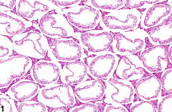
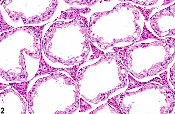
生精上皮萎缩(Germinal epithelium atrophy)(Figure 1、Figure 2、Figure 3、Figure 4)表现为生精小管完全缺乏生殖细胞,仅由支持细胞(Sertoli cell)衬覆。生精小管可出现管腔扩张(Figure 1、Figure 2)或管腔收缩(Figure 4)。若管腔扩张,萎缩很可能由长期严重的管腔扩张所致压力(即压迫性萎缩)引起(另见 Testis - Seminiferous Tubule - Dilation)。该病变可为单侧或双侧,呈 局灶性(focal)、多灶性(multifocal) 或 弥漫性(diffuse),在小鼠和大鼠各年龄段均可作为偶发性发现出现,发生率随年龄增长而升高。生精上皮萎缩是一种终末期病变,通常先有或伴有生精小管变性。依严重程度不同,受累睾丸肉眼观可松弛,体积和重量减小。严重的 弥漫性 生精上皮萎缩常不可逆。大鼠中可见散在生精小管伴生殖细胞萎缩,属衰老性改变。
诊断与分级建议(NTP 共识)
若生精上皮萎缩被认为是主要病变,且其发生率和/或严重程度似乎与受试物给药相关,则应作出诊断、进行分级,并在病理学叙述中加以讨论。由于生精上皮萎缩被视为变性的终末期,萎缩的诊断应保留用于大多数受累小管缺乏生殖细胞的病例,否则变性是更合适的诊断(见 Testis - Germinal Epithelium - Degeneration)。若同时存在两种病变(既有萎缩的小管又有变性小管),可在病理学叙述中描述。若同一研究中同时使用这两个术语,病理学家有责任在病理学叙述中描述两种病变之间的关系。若双侧睾丸均受累,诊断应标明为双侧,严重程度分级应基于受累更严重的睾丸。相关链接:Testis - Seminiferous Tubule - Dilation;Testis - Germinal Epithelium - Degeneration
图谱
英文原文
展开 NTP 官方英文描述与建议
Comment:
Germinal epithelium atrophy ( Figure 1 , Figure 2 , Figure 3 , and Figure 4 ) consists of seminiferous tubules that are completely devoid of germ cells and lined only by Sertoli cells. The seminiferous tubules may have dilated lumens ( Figure 1 and Figure 2 ) or contracted lumens ( Figure 4 ). If the tubular lumens are dilated, the atrophy is likely caused by pressure (i.e. pressure atrophy) fromprolonged and severe tubular dilation (also see Testis - Seminiferous Tubule - Dilation ) The change may be unilateral or bilateral and focal, multifocal, or diffuse and occurs as an occasional incidental finding in mice and rats at all ages, with the incidence increasing with age. Germinal epithelial atrophy is an end-stage lesion and is generally preceded, or accompanied by, seminiferous tubule degeneration. Depending on severity, the affected testes may be macroscopically flaccid and reduced in size and weight. Severe, diffuse germinal epithelial atrophy is often irreversible. Scattered seminiferous tubules with germinal cell atrophy can be seen as an aging change in rats.
Recommendation:
Germinal epithelial atrophy should be diagnosed, graded and should be discussed in the pathology narrative if it is considered the primary change and if the incidence and/or severity appears to be related to chemical administration. Since germinal epithelial atrophy is considered the end stage of degeneration, the diagnosis of atrophy should be reserved for cases in which the majority of the affected tubules are devoid of germ cells, otherwise, degeneration is a more appropriate diagnosis (see Testis - Germinal Epithelium - Degeneration ). If there is a mixture of effects (both atrophied and degenerative tubules), it can be described in the pathology narrative. If both terms are used in a single study, it is incumbent upon the pathologist to describe the relationship between the two lesions in the pathology narrative. If both testes are affected, the diagnosis should be indicated as bilateral and the severity grade based on the more severely affected testis. Related Links: Testis - Seminiferous Tubule - Dilation Testis - Germinal Epithelium - Degeneration
来源与引用
原始条目:https://ntp.niehs.nih.gov/atlas/nnl/reproductive-system-male/testis/GerminalEpithelium-Atrophy
图谱总目录:NTP Nonneoplastic Lesion Atlas。 内容属美国联邦政府作品(公有领域),可引用;引用时请标注来源。
建议引用格式:NTP Nonneoplastic Lesion Atlas. Germinal Epithelium Atrophy (testis). LabSmart 中文版:
https://www.labsmart.cn/ntp/testis-germinal-epithelium-atrophy.html
中文图谱总页(可展开对照全部 595 条):https://www.labsmart.cn/ntp-atlas.html#testis-germinal-epithelium-atrophy