生精小管巨细胞Seminiferous Tubule Giant Cells · 睾丸 · 雄性生殖系统 · NTP ntp-0464
诊断要点:当多核巨细胞构成睾丸病变中生精细胞退变的主要形式时,应当诊断并分级。存在双侧累及时应当记录,因为单侧性常提示为偶然性病变。
病变描述
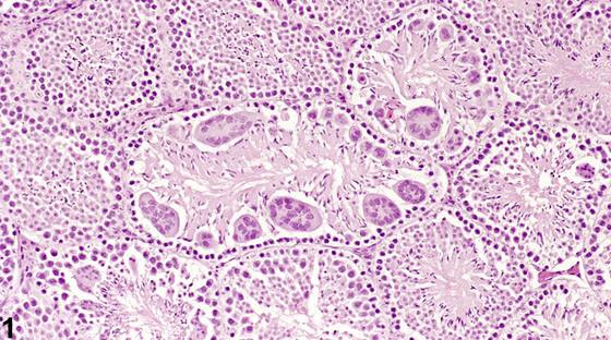
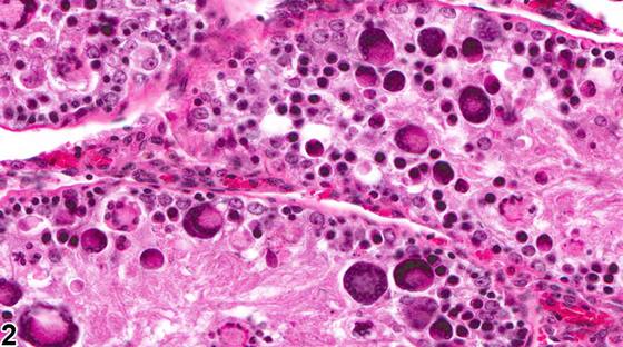
生精小管内的多核巨细胞是退变生精细胞的一种特定形式,与非特异性生精细胞退变不同(见睾丸-生精上皮-退变)。多核巨细胞的存在可为局灶性(focal)或弥漫性(diffuse),并可累及一侧或双侧睾丸。多核巨细胞偶尔可作为背景性偶然病变出现。该现象的发生是因为正常生精细胞分裂的特征是不完全胞质分裂,因此每次细胞分裂的子代细胞通过胞质桥相互连接。在某些形式的退变过程中,这些胞质桥可以开放,使相连细胞的胞质内容物融合。一定程度的生精细胞退变/萎缩可伴有多核巨细胞形成,若属此种情况,则不应将其作为一项独立所见记录。然而,如果多核巨细胞作为睾丸病变的主要特征存在,如Figure 1、Figure 2所示,则记录“多核巨细胞”这一所见可能是恰当的。
诊断与分级建议(NTP 共识)
当多核巨细胞构成睾丸病变中生精细胞退变的主要形式时,应当诊断并分级。存在双侧累及时应当记录,因为单侧性常提示为偶然性病变。如果多核巨细胞仅作为非特异性小管退变的次要伴随现象出现,则不应将其作为一项独立所见记录,但可在病理学描述中讨论。相关链接:睾丸-生精上皮-退变
图谱
英文原文
展开 NTP 官方英文描述与建议
Comment:
Multinucleated giant cells in seminiferous tubules are a specific form of degenerating germ cell, in contrast to nonspecific germ cell degeneration (see Testis - Germinal Epithelium - Degeneration ). The presence of giant cells can be focal or diffuse and may affect one or both testes. Multinucleated giant cells can occasionally be seen as an incidental background lesion. The phenomenon occurs because normal germ cell division is characterized by incomplete cytokinesis, so the progeny of each cell division are joined to each other by cytoplasmic bridges. During some forms of degeneration, these cytoplasmic bridges can open and allow fusion of the cellular contents of the conjoined cells. Some degree of multinucleated giant cell formation may accompany germinal cell degeneration/atrophy, and if this is the case, they should not be recorded as a separate finding. However, if multinucleated giant cells are present as a dominant feature of a testicular lesion, as in Figure 1 and Figure 2 , the finding of “multinucleated giant cells” may be warranted.
Recommendation:
Multinucleated giant cells should be diagnosed and graded when they represent the predominant form of germ cell degeneration in a testicular lesion. Bilateral involvement should be recorded when present because unilaterality frequently provides evidence of an incidental lesion. If multinucleated giant cells occur as a minor accompaniment to nonspecific tubular degeneration, they should not be recorded as a separate finding but may be discussed in the pathology narrative. Related Links: Testis - Germinal Epithelium - Degeneration
来源与引用
原始条目:https://ntp.niehs.nih.gov/atlas/nnl/reproductive-system-male/testis/SeminiferousTubule-GiantCells
图谱总目录:NTP Nonneoplastic Lesion Atlas。 内容属美国联邦政府作品(公有领域),可引用;引用时请标注来源。
建议引用格式:NTP Nonneoplastic Lesion Atlas. Seminiferous Tubule Giant Cells (testis). LabSmart 中文版:
https://www.labsmart.cn/ntp/testis-seminiferous-tubule-giant-cells.html
中文图谱总页(可展开对照全部 595 条):https://www.labsmart.cn/ntp-atlas.html#testis-seminiferous-tubule-giant-cells