生精小管坏死Seminiferous Tubule Necrosis · 睾丸 · 雄性生殖系统 · NTP ntp-0463
诊断要点:生精小管坏死应当诊断并分级,并且如果其发生率和/或严重程度似乎与给予化学物质相关,应当在病理学描述中加以讨论。
病变描述
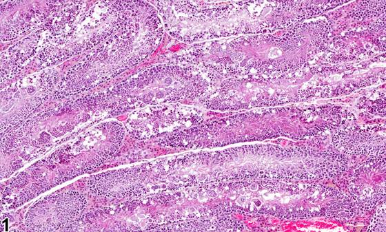
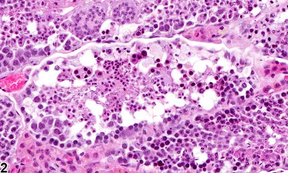
生精小管坏死(seminiferous tubule necrosis)(Figure 1、Figure 2)的特征是生精小管内的生殖细胞和支持细胞发生细胞死亡。Figure 1、Figure 2中的细胞死亡由以下表现构成:细胞呈强嗜酸性、排列紊乱,可见核固缩并伴有空泡化,或继发于缺血或梗死的大片淡嗜酸性蛋白凝固区(Figure 3、Figure 4)。生精小管坏死最常见的原因是缺血性损伤,可能源于睾丸的偶然性扭转,也可能与化学物质相关。此外,一些血管活性物质,如5-羟色胺、组胺和肾上腺素,也已被证明可导致生精小管坏死。这种细胞死亡形式的一个显著特征是同时累及生精上皮和支持细胞。相比之下,仅累及生精上皮的细胞死亡通常通过细胞凋亡过程发生。已有报道,给予啮齿类动物镉后可发生化学诱导的睾丸坏死,镉是啮齿类动物睾丸的内皮毒物。坏死也可由引起睾丸血管血栓形成的物质所致。有报道称,给大鼠单次给予人绒毛膜促性腺激素后,大鼠睾丸前下部出现特征性的局灶性(focal)坏死(Figure 4)。该坏死被认为是由于Leydig细胞释放前列腺素引起的局部缺血所致。睾丸坏死不可逆,原因是支持细胞和管状结构丧失,受累区域被瘢痕组织取代。睾丸的大范围坏死也可由意外将药物直接腹腔内注射入睾丸所致(这种情况可能发生,因为啮齿类动物能将睾丸回缩入腹腔)。
诊断与分级建议(NTP 共识)
生精小管坏死应当诊断并分级,并且如果其发生率和/或严重程度似乎与给予化学物质相关,应当在病理学描述中加以讨论。如果双侧睾丸均受累,诊断应当限定为双侧(bilateral),并根据受累更严重的睾丸给出严重程度分级。如果血管血栓形成是明显的促发原因,该事实应当在病理学描述中提及。相关病变,如炎症,如果严重程度足以需要,应当单独诊断。
图谱
英文原文
展开 NTP 官方英文描述与建议
Comment:
Seminiferous tubule necrosis ( Figure 1 and Figure 2 ) is characterized by seminiferous tubules in which germ cells and Sertoli cells are undergoing cell death. The cell death in Figure 1 and Figure 2 consists of hypereosinophilic disorganized cells showing pyknosis and associated vacuolization or larger areas of pale eosinophilic regions of protein coagulation secondary to ischemia or infarction ( Figure 3 and Figure 4 ). The most common cause of seminiferous tubule necrosis is ischemic injury, which may result from incidental torsion of the testis or may be chemically related. In addition, some vasoactive agents, such as serotonin, histamine, and epinephrine, have also been shown to produce seminiferous tubule necrosis. A salient feature of this form of cell death is that it involves both germinal epithelium and Sertoli cells. In contrast, cell death involving only germinal epithelium typically occurs through the process of apoptosis. Chemically induced testicular necrosis has been described in rodents administered cadmium, which is an endothelial toxicant in the testis of rodents. Necrosis can also be caused by agents causing vascular thrombosis in the testis. A characteristically focal necrosis at the frontal lower part of the rat testis ( Figure 4 ) has been described following a single administration of human chorionic gonadotropin to rats. The necrosis was considered to be due to local ischemia caused by prostaglandin release from Leydig cells. Testicular necrosis is not reversible, due to loss of Sertoli cells and tubular structure, and the affected area is replaced by scar tissue. Extensive necrosis of the testis can also be caused by accidental intraperitoneal injection directly into the testis (which can occur because the rodent is able to retract its testes into the abdominal cavity).
Recommendation:
Seminiferous tubule necrosis should be diagnosed and graded and should be discussed in the pathology narrative if the incidence and/or severity appear to be related to chemical administration. If both testes are affected, the diagnosis should be qualified as bilateral and given a severity grade based on the more severely affected testis. If vascular thrombi are evident as a precipitating cause, that fact should be mentioned in the pathology narrative. Associated lesions, such as inflammation, should be diagnosed separately if warranted by severity.
来源与引用
原始条目:https://ntp.niehs.nih.gov/atlas/nnl/reproductive-system-male/testis/SeminiferousTubule-Necrosis
图谱总目录:NTP Nonneoplastic Lesion Atlas。 内容属美国联邦政府作品(公有领域),可引用;引用时请标注来源。
建议引用格式:NTP Nonneoplastic Lesion Atlas. Seminiferous Tubule Necrosis (testis). LabSmart 中文版:
https://www.labsmart.cn/ntp/testis-seminiferous-tubule-necrosis.html
中文图谱总页(可展开对照全部 595 条):https://www.labsmart.cn/ntp-atlas.html#testis-seminiferous-tubule-necrosis