间质细胞增生Interstitial Cell Hyperplasia · 睾丸 · 雄性生殖系统 · NTP ntp-0477
诊断要点:间质细胞增生若因处理而加重,应作出诊断、分级并在病理学叙述中讨论。当双侧睾丸均存在时,诊断应指明该病变为双侧性,严重程度应基于受累更严重的睾丸。
病变描述
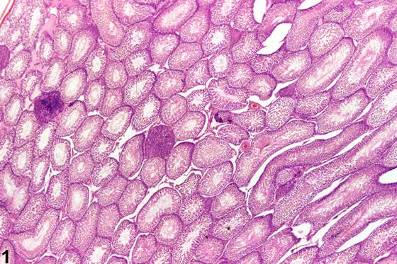
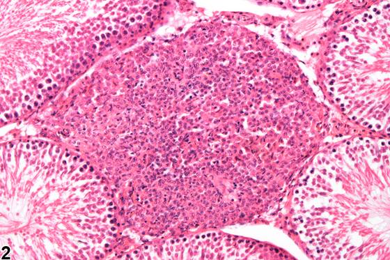
间质细胞(Leydig细胞)增生可呈局灶性(focal)、多灶性(multifocal)(Figure 1、Figure 2)或弥漫性(diffuse)(Figure 3、Figure 4),由间质细胞数量增多构成,对邻近生精小管无压迫性。弥漫性增生通常是激素失衡的生理性反应,而局灶性增生常构成通向间质细胞腺瘤的连续病变过程的一部分。局灶性增生与间质细胞腺瘤的区分常基于大小,增生被定义为直径等于或小于三个生精小管。该病变可与睾丸萎缩相关。间质细胞的增殖性病变是常见的年龄相关睾丸病变,在F344大鼠中尤为常见,但在Wistar大鼠和Sprague-Dawley大鼠中也有较低发生率。小鼠中也存在品系差异。在大鼠中,任何升高循环黄体生成素水平的化学物均可轻易诱导间质细胞增生和腺瘤。这包括许多不同结构类别的化学物和药物。在小鼠中,雌激素类化合物是间质细胞增殖最常见的原因。
诊断与分级建议(NTP 共识)
间质细胞增生若因处理而加重,应作出诊断、分级并在病理学叙述中讨论。当双侧睾丸均存在时,诊断应指明该病变为双侧性,严重程度应基于受累更严重的睾丸。
图谱
英文原文
展开 NTP 官方英文描述与建议
Comment:
Hyperplasia of interstitial cells (Leydig cells) can be focal, multifocal ( Figure 1 and Figure 2 ), or diffuse ( Figure 3 and Figure 4 ) and consists of an increased number of interstitial cells, which are noncompressive to the adjacent seminiferous tubules. Diffuse hyperplasia is generally a physiologic response to hormonal imbalance, whereas focal hyperplasia commonly forms part of the continuum leading to interstitial cell adenoma. Distinction between focal hyperplasia and interstitial cell adenoma is frequently based on size, with hyperplasia being defined as having a diameter equal to three or fewer seminiferous tubules. The condition may be associated with testicular atrophy. Proliferative lesions of the interstitial cell are common age-related lesions of the testis, being particularly frequent in the F344 rat but also present at lower incidences in the Wistar rat and Sprague-Dawley rat. Strain differences are also present in mice. Interstitial cell hyperplasia and adenoma can be readily induced in rats by any chemical that increases circulating levels of luteinizing hormone. This includes many different structural classes of chemicals and drugs. In mice, estrogenic compounds are the most common cause of interstitial cell proliferation.
Recommendation:
Interstitial cell hyperplasia should be diagnosed, graded, and discussed in the pathology narrative, if exacerbated by treatment. When present in both testes, the diagnosis should indicate the condition is bilateral, and severity should be based on the more severely affected testis.
来源与引用
原始条目:https://ntp.niehs.nih.gov/atlas/nnl/reproductive-system-male/testis/InterstitialCell-Hyperplasia
图谱总目录:NTP Nonneoplastic Lesion Atlas。 内容属美国联邦政府作品(公有领域),可引用;引用时请标注来源。
建议引用格式:NTP Nonneoplastic Lesion Atlas. Interstitial Cell Hyperplasia (testis). LabSmart 中文版:
https://www.labsmart.cn/ntp/testis-interstitial-cell-hyperplasia.html
中文图谱总页(可展开对照全部 595 条):https://www.labsmart.cn/ntp-atlas.html#testis-interstitial-cell-hyperplasia