睾丸网增生Rete Hyperplasia · 睾丸 · 雄性生殖系统 · NTP ntp-0468
诊断要点:睾丸网增生只要存在就应诊断并分级。存在双侧受累时应记录。相关病变如精子淤滞和生精小管扩张除非因严重程度需要,否则不应单独诊断,但应在病理叙述中描述。
病变描述
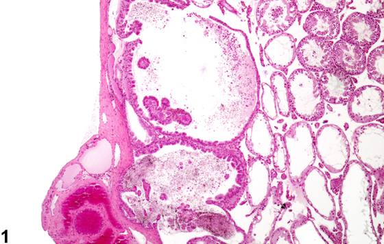
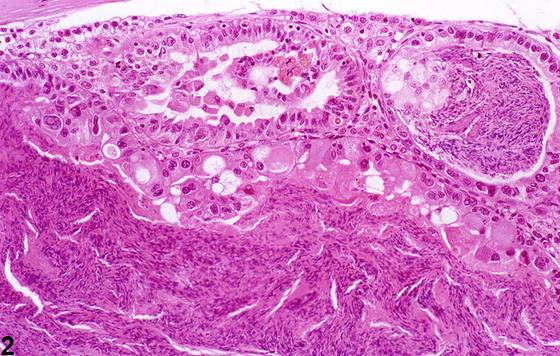
睾丸网(rete testis)上皮增生以局灶性(focal)或多灶性(multifocal)病变形式发生,在大鼠中可作为偶发性病变见到,但在CD1小鼠中更为常见。增生的上皮一般向管腔内形成乳头状或结节状突起,不压迫周围组织(Figure 1、Figure 2)。该增生常伴有睾丸网管囊性扩张和精子淤滞(Figure 2)。已观察到睾丸网增殖性病变是对产前暴露于己烯雌酚(diethyl stilbestrol)的反应。该发现需与睾丸网腺瘤(rete testis adenoma)相鉴别,后者广泛的乳头状结构和支持性间质会压迫周围组织。
诊断与分级建议(NTP 共识)
睾丸网增生只要存在就应诊断并分级。存在双侧受累时应记录。相关病变如精子淤滞和生精小管扩张除非因严重程度需要,否则不应单独诊断,但应在病理叙述中描述。
图谱
英文原文
展开 NTP 官方英文描述与建议
Comment:
Hyperplasia of the rete testis epithelium occurs as a focal or multifocal lesion and can be seen as an occasional incidental lesion in rats but more commonly in CD1 mice. The hyperplastic epithelium generally forms papillary or knob-like projections into the lumen with no compression of surrounding tissue ( Figure 1 and Figure 2 ). The hyperplasia is frequently accompanied by cystic dilation of the rete ducts and sperm stasis ( Figure 2 ). Proliferative lesions of the rete testis have been observed in response to prenatal exposure to diethyl stilbestrol. The finding needs to be distinguished from rete testis adenoma, where extensive papillary structures and supporting stroma cause compression of the surrounding tissue.
Recommendation:
Rete testis hyperplasia should be diagnosed and graded whenever present. Bilateral involvement should be recorded when present. Associated lesions such as sperm stasis and seminiferous tubule dilation should not be diagnosed separately unless warranted by severity, but should be described in the pathology narrative.
来源与引用
原始条目:https://ntp.niehs.nih.gov/atlas/nnl/reproductive-system-male/testis/ReteHyperplasia
图谱总目录:NTP Nonneoplastic Lesion Atlas。 内容属美国联邦政府作品(公有领域),可引用;引用时请标注来源。
建议引用格式:NTP Nonneoplastic Lesion Atlas. Rete Hyperplasia (testis). LabSmart 中文版:
https://www.labsmart.cn/ntp/testis-rete-hyperplasia.html
中文图谱总页(可展开对照全部 595 条):https://www.labsmart.cn/ntp-atlas.html#testis-rete-hyperplasia