生精小管扩张Seminiferous Tubule Dilation · 睾丸 · 雄性生殖系统 · NTP ntp-0465
诊断要点:小管扩张应当被诊断并分级,并且如果其发生率或严重程度似乎与化学物给药相关,应当在病理学叙述中加以讨论。
病变描述
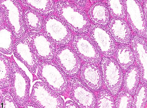
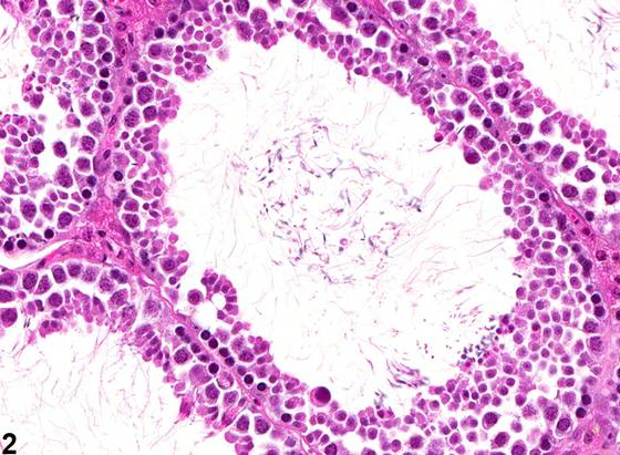
生精小管扩张(seminiferous tubule dilation)的特征是生精小管管腔直径增大(Figure 1、Figure 2)。这与正常睾丸生精小管中不明显的管腔空间形成对比(Figure 3、Figure 4)。受累小管的生精上皮变薄并受压,这是由管腔内生精小管液体积增加所致的压力积聚引起的。液体增加可能是由于输出小管(efferent ducts)对液体的重吸收减少、输出小管阻塞、生精小管液体排空减少,或Sertoli细胞产生生精小管液体增加。如果液体压力持续存在,小管扩张可迅速进展为严重的生精上皮萎缩,导致生精过程不可逆地停止。当小管扩张继发于输出小管阻塞时,病变可能为单侧,而阻塞的原因仍可能与受试物相关。除非对输出小管进行采样(这在啮齿类动物中较为困难),否则小管扩张的原因将不明显。ERKO小鼠在达到青春期时发生生精小管扩张,随后出现严重的生精上皮萎缩。这是输出小管未能重吸收生精小管液体的继发后果,而该重吸收是一种雌激素依赖性功能。已有报道多种化学物可引起生精小管扩张及其萎缩后遗症,包括杀菌剂多菌灵(carbendazim)、一种PDE4抑制剂、一种白三烯抑制剂和内皮素拮抗剂。
诊断与分级建议(NTP 共识)
小管扩张应当被诊断并分级,并且如果其发生率或严重程度似乎与化学物给药相关,应当在病理学叙述中加以讨论。当存在时,应当记录双侧受累,因为单侧性通常反映对输出小管病变的继发反应。还应当注意单侧或双侧生精小管萎缩的发生率,因为这可能是小管扩张的进展性和终末期病变。还应当注意附睾起始段或附睾脂肪垫(输出小管所在位置)内任何大体或显微镜下病变的证据,这些病变可能反映精子肉芽肿、精子淤滞或扩张的管道,这将提示起始性、阻塞性病变的可能来源。
图谱
英文原文
展开 NTP 官方英文描述与建议
Comment:
Seminiferous tubule dilation is characterized by increased luminal diameter of the seminiferous tubules ( Figure 1 and Figure 2 ). This contrasts with the inconspicuous luminal space in the seminiferous tubules from a normal testis ( Figure 3 and Figure 4 ). The affected tubules have thinning and compression of the seminiferous epithelium, which is caused by pressure buildup from increased volume of seminiferous tubule fluid within the lumen. The increased fluid can be due to decreased reabsorption of fluid in the efferent ducts, efferent duct blockage, decreased emptying of fluid from the seminiferous tubules, or increased production of seminiferous tubule fluid by the Sertoli cells. If the fluid pressure is maintained, the tubular dilation can rapidly progress to severe germinal epithelial atrophy, resulting in an irreversible cessation of spermatogenesis. When the tubular dilation is secondary to blockage of the efferent ducts, the lesion may be unilateral, and yet the cause of the blockage can still be test article related. Unless the efferent ducts are sampled (which is difficult in rodents), the cause of the tubular dilation will not be apparent. The ERKO mouse develops seminiferous tubular dilation followed by severe germinal epithelial atrophy as it reaches puberty. This occurs as a secondary consequence to the failure of the efferent ducts to resorb seminiferous tubule fluid, which is an estrogen-dependent function. Seminiferous tubular dilatation with its sequel of atrophy has been reported for a number of chemicals, including the fungicide carbendazim, a PDE4 inhibitor, a leukotriene inhibitor, and endothelin antagonists.
Recommendation:
Tubular dilatation should be diagnosed and graded and should be discussed in the pathology narrative if the incidence and/or severity appears to be related to chemical administration. When present, bilateral involvement should be recorded because unilaterality usually reflects a secondary response to lesions in the efferent ducts. Attention should also be paid to the incidence of unilateral or bilateral seminiferous tubule atrophy since this can be a progressive and end-stage lesion of tubular dilation. Attention should also be paid to any evidence of gross or microscopic lesions within the initial segment of the epididymis or the epididymal fat pad (location of the efferent ducts) that may reflect sperm granulomas, sperm stasis, or dilated ducts, which would indicate the likely origin of the initiating, obstructive lesion.
来源与引用
原始条目:https://ntp.niehs.nih.gov/atlas/nnl/reproductive-system-male/testis/SeminiferousTubule-Dilation
图谱总目录:NTP Nonneoplastic Lesion Atlas。 内容属美国联邦政府作品(公有领域),可引用;引用时请标注来源。
建议引用格式:NTP Nonneoplastic Lesion Atlas. Seminiferous Tubule Dilation (testis). LabSmart 中文版:
https://www.labsmart.cn/ntp/testis-seminiferous-tubule-dilation.html
中文图谱总页(可展开对照全部 595 条):https://www.labsmart.cn/ntp-atlas.html#testis-seminiferous-tubule-dilation