生精小管空泡化Seminiferous Tubule Vacuolation · 睾丸 · 雄性生殖系统 · NTP ntp-0466
诊断要点:生精小管空泡化应当进行诊断和分级,并且如果其发生率和/或严重程度似乎与受试物给药相关,则应当在病理学叙述中加以讨论。
病变描述
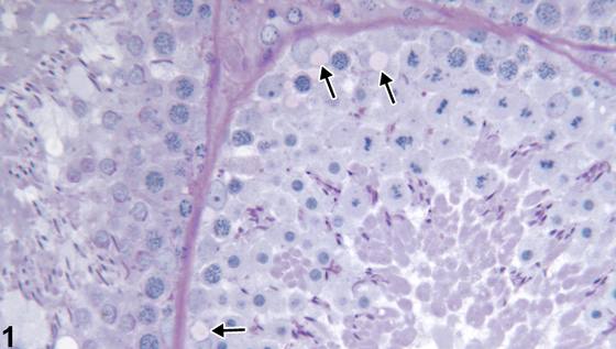
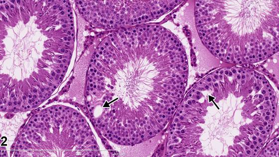
生精小管空泡化(seminiferous tubule vacuolation)在形态上可表现为微空泡或大空泡。微空泡化通常发生于基底部的Sertoli细胞胞质内(Figure 1,箭头),而大空泡化则表现为生精上皮深部出现一个或几个空泡(Figure 2,箭头)。在大多数病例中,空泡位于Sertoli细胞内或Sertoli细胞之间。空泡内可能含有液体或脂质,也可能代表内质网或细胞间隙的扩张。超微结构检查有助于确定其来源。Sertoli细胞空泡化是Sertoli细胞形态学损伤常见的早期特征,在为期28天或更短时间的研究中即可见到,且出现在任何生殖细胞变性之前。另外,空泡常伴随弥漫性生殖细胞变性/萎缩出现,此时空泡可能代表缺失的生殖细胞所留下的空隙。在对照睾丸中,空泡常可见于偶见小管的偶然性发现(Figure 2,箭头),并且其发生率和/或严重程度可能随时间推移而增加。
诊断与分级建议(NTP 共识)
生精小管空泡化应当进行诊断和分级,并且如果其发生率和/或严重程度似乎与受试物给药相关,则应当在病理学叙述中加以讨论。如果双侧睾丸均出现空泡化,则应当记录为双侧性,严重程度分级应基于受累更严重的睾丸。
图谱
英文原文
展开 NTP 官方英文描述与建议
Comment:
Seminiferous tubule vacuolation can be microvacuolar or macrovacuolar in appearance. Microvacuolation generally occurs in the basal Sertoli cell cytoplasm ( Figure 1 , arrows), and macrovacuolation appears as one or a few vacuoles within the depth of the seminiferous epithelium ( Figure 2 , arrows). In most cases, the vacuoles are within or between Sertoli cells. They may contain fluid or lipid, or they may represent dilation of endoplasmic reticulum or intercellular spaces. Ultrastructural studies can be helpful to determine their origin. Vacuolation of Sertoli cells is a common early feature of morphologic injury to Sertoli cells that may be seen in studies of 28 days or less duration, prior to any germ cell degeneration. Alternatively, vacuoles often accompany generalized germinal cell degeneration/atrophy, where they probably represent spaces left by the missing germ cells. Vacuoles can often be seen as an incidental finding in occasional tubules in control testes ( Figure 2 , arrows) and may increase in incidence and/or severity over time.
Recommendation:
Seminiferous tubule vacuolation should be diagnosed and graded and should be discussed in the pathology narrative if the incidence and/or severity appears to be related to chemical administration. If present in both testes, the vacuolation should be recorded as bilateral and the severity grade based on the more severely affected testis.
来源与引用
原始条目:https://ntp.niehs.nih.gov/atlas/nnl/reproductive-system-male/testis/SeminiferousTubule-Vacuolation
图谱总目录:NTP Nonneoplastic Lesion Atlas。 内容属美国联邦政府作品(公有领域),可引用;引用时请标注来源。
建议引用格式:NTP Nonneoplastic Lesion Atlas. Seminiferous Tubule Vacuolation (testis). LabSmart 中文版:
https://www.labsmart.cn/ntp/testis-seminiferous-tubule-vacuolation.html
中文图谱总页(可展开对照全部 595 条):https://www.labsmart.cn/ntp-atlas.html#testis-seminiferous-tubule-vacuolation