扩张Dilation · 子宫 · 雌性生殖系统 · NTP ntp-0535
诊断要点:Uterus - Dilation 当不属于正常发情周期性变化时,应记录并分级。
病变描述
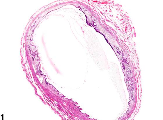
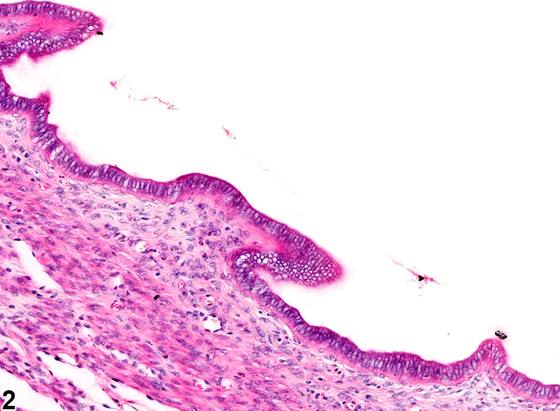
子宫角扩张(Figure 1、Figure 2、Figure 3、Figure 4)在剖检时常见,且这些子宫内常在腔内蓄积过量液体。子宫扩张在 rat 和 mouse 中均相对常见,并可能呈节段性。腔性扩张可能与间质息肉(stromal polyp)相关,或继发于卵巢囊肿所致激素失衡,或继发于老年啮齿类动物发情周期停止后的持续发情状态。在 rat 中给予孕激素(progestin)、雌激素(estrogen)和他莫昔芬(tamoxifen)与子宫扩张相关。在周期性发情的啮齿类动物中,腔性扩张通常在发情前期(proestrus)和发情期(estrus)可见,不应作出诊断。浆液性液体产生增加是发情周期发情前期的一部分,依据阴道上皮判断(其显示早期角化,表面覆盖一层黏液化细胞),不应作出诊断。子宫扩张时,子宫内膜衬里通常变薄或萎缩,且由于压力增加,子宫壁变薄,但在较轻病例中子宫内膜可正常(Figure 2)。继发于炎症过程(如子宫积脓,pyometra)的扩张子宫腔不应单独诊断。子宫扩张可能需要与 显著(marked) 囊性子宫内膜增生(cystic endometrial hyperplasia)相鉴别,尤其是仅检查子宫横切面时。在囊性子宫内膜增生中,活跃增殖的腺体数量增加,其腔可能扩张。在子宫扩张中,子宫腔扩张,可能与单个扩张的腺腔混淆。因此,必须仔细检查切片,以确定扩张结构是单个子宫腺还是子宫腔。这两种病变的鉴别在子宫纵切面中更为明显。
诊断与分级建议(NTP 共识)
Uterus - Dilation 当不属于正常发情周期性变化时,应记录并分级。
图谱
英文原文
展开 NTP 官方英文描述与建议
Comment:
Dilation of uterine horns ( Figure 1 , Figure 2 , Figure 3 , and Figure 4 ) is commonly observed at necropsy, and frequently these uteri have accumulations of excessive amounts of fluid within the lumen. Uterine dilation is relatively commonly seen in both rats and mice and may be segmental. Luminal dilation may be associated with stromal polyps or occur secondarily to hormonal imbalances from ovarian cysts or to a prolonged estrus state after cessation of the estrus cycle in aged rodents. Administration of progestins, estrogens, and tamoxifen in rats has been associated with uterine dilation. Luminal dilation is normally observed at proestrus and estrus in cycling rodents and should not be diagnosed. Increased serous fluid production is part of the proestrus phase of the cycle judged by the vaginal epithelium (which shows early keratinization covered by a layer of mucified cells) and should not be diagnosed. With uterine dilation, the endometrial lining is usually attenuated or atrophic and the wall of the uterus thinned due to the increasing pressure, but in less severe cases the endometrium can be normal ( Figure 2 ). Dilated uterine lumen that is secondary to an inflammatory process such as pyometra should not be diagnosed separately. Uterine dilation may need to be differentiated from marked cystic endometrial hyperplasia, particularly when only cross-sections of the uterus are examined. In cystic endometrial hyperplasia, there is an increased number of active proliferating glands that may have dilated lumens. In uterine dilation, the uterine lumen is dilated, which could be confused with a single dilated glandular lumen. Therefore, the section must be carefully examined to determine whether the dilated structure is a single uterine gland or is the uterine lumen. The differentiation of these two lesions is more obvious in longitudinal sections of uterus.
Recommendation:
Uterus - Dilation should be recorded and graded when not part of normal estrous cyclicity.
来源与引用
原始条目:https://ntp.niehs.nih.gov/atlas/nnl/reproductive-system-female/uterus/Dilation
图谱总目录:NTP Nonneoplastic Lesion Atlas。 内容属美国联邦政府作品(公有领域),可引用;引用时请标注来源。
建议引用格式:NTP Nonneoplastic Lesion Atlas. Dilation (uterus). LabSmart 中文版:
https://www.labsmart.cn/ntp/uterus-dilation.html
中文图谱总页(可展开对照全部 595 条):https://www.labsmart.cn/ntp-atlas.html#uterus-dilation