上皮增生Epithelium Hyperplasia · 阴道 · 雌性生殖系统 · NTP ntp-0558
诊断要点:阴道,上皮——增生:当与处理相关时,应作出诊断并分级。如果上皮增厚是与正常动情周期相关的正常变化,则不应诊断为增生。
病变描述
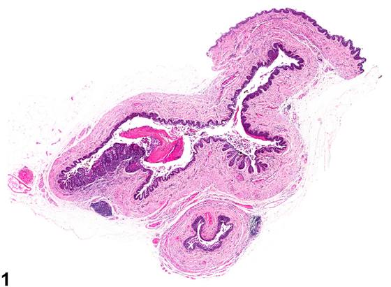
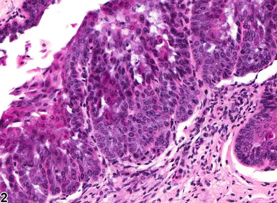
啮齿类阴道上皮增生(Figure 1、Figure 2、Figure 3、Figure 4)常伴有过角化(hyperkeratosis),且通常为弥漫性。细胞分化良好,无异型性,也无侵袭下方间质的表现。增生的细胞可向下方间质形成向下突起(网嵴,rete pegs)(Figure 3、Figure 4)。增生可发生于多种情形,包括生殖衰老期处于持续动情期的对照大鼠,以及继发于内源性雌二醇生成增加或给予具有激素活性的外源化学物。阴道上皮的局灶性(focal)增生不常见,但已有观察到。增生应与动情周期中阴道外观的正常变异相鉴别(周期性变化的描述见引言)。增生是阴道上皮厚度的增加(与正常 cycling 动物的正常动情期相比)。局灶性增生的鉴别诊断包括乳头状瘤(papilloma)和息肉(polyp)。增生细胞明显位于上皮基底膜之上,不形成离散的上皮细胞增殖性团块。增生局限于正常黏膜表面,不突入管腔。息肉主要由被覆正常或轻度角化上皮的纤维肌性核心构成。
诊断与分级建议(NTP 共识)
阴道,上皮——增生:当与处理相关时,应作出诊断并分级。如果上皮增厚是与正常动情周期相关的正常变化,则不应诊断为增生。与增生相关的过角化除非严重程度确有必要,否则不应单独诊断,但应在病理学描述中予以描述。
图谱
英文原文
展开 NTP 官方英文描述与建议
Comment:
Hyperplasia of the rodent vaginal epithelium ( Figure 1 , Figure 2 , Figure 3 and Figure 4 ) is often associated with hyperkeratosis and is usually diffuse. Cells are well differentiated with no atypia or invasion of the underlying stroma. Hyperplastic cells may form downward projections into the underlying stroma (rete pegs) ( Figure 3 and Figure 4 ). Hyperplasia occurs in a variety of settings, including control rats in persistent estrus during reproductive senescence, and secondary to increases in endogenous estradiol production or administration of hormonally active xenobiotics. Focal hyperplasia of vaginal epithelium is uncommon but has been observed. Hyperplasia should be differentiated from normal variation in the appearance of the vagina during the estrus cycle (see Introduction for description of cyclical changes). Hyperplasia is increased thickness of the vaginal epithelium (compared with normal estrus in a normally cycling animal). The differential diagnoses for focal hyperplasia are papilloma and polyp. Hyperplastic cells are evident above the basement membrane of the epithelium and do not form a discrete proliferative mass of epithelial cells. Hyperplasia is restricted to the normal mucosal surface and does not protrude into the lumen. A polyp is composed predominantly of a fibromuscular core covered by normal or slightly hyperkeratinized epithelium.
Recommendation:
Vagina, Epithelium - Hyperplasia should be diagnosed and graded when it is treatment related. If the epithelial thickening is a normal change associated with the normal estrous cycle, it should not be diagnosed as hyperplasia. Hyperkeratosis associated with the hyperplasia should not be diagnosed separately unless warranted by severity but should be described in the pathology narrative.
来源与引用
原始条目:https://ntp.niehs.nih.gov/atlas/nnl/reproductive-system-female/vagina/Epithelium-Hyperplasia
图谱总目录:NTP Nonneoplastic Lesion Atlas。 内容属美国联邦政府作品(公有领域),可引用;引用时请标注来源。
建议引用格式:NTP Nonneoplastic Lesion Atlas. Epithelium Hyperplasia (vagina). LabSmart 中文版:
https://www.labsmart.cn/ntp/vagina-epithelium-hyperplasia.html
中文图谱总页(可展开对照全部 595 条):https://www.labsmart.cn/ntp-atlas.html#vagina-epithelium-hyperplasia