精子减少Hypospermia · 附睾 · 雄性生殖系统 · NTP ntp-0502
诊断要点:精子含量减少应记录并分级,并且如果其发生率和/或严重程度似乎与化学物给药相关,则应在病理学叙述中加以讨论。
病变描述
精子含量减少通常由睾丸精子发生减少所致,但也可能源于输出管中的阻塞性病变,使外观正常的睾丸产生的精子无法到达附睾。精子通常在3-5天内到达附睾尾并在该处储存一周或更长时间。某些毒物可导致转运时间缩短,从而减少正常精子含量(而对精子发生无影响)。睾酮水平降低也会导致附睾中精子存活率下降。精子减少常伴有脱落的睾丸生精细胞和细胞碎片增多,这是由于睾丸中相关的精子发生障碍所致。精子含量减少还常与精子及液体体积减少所致的管腔缩小(导管萎缩)相关。精子可能减少(少精子症,oligospermia)或完全缺失(无精子症,aspermia);减少的程度可用严重程度分级来表示。围青春期大鼠(8-10周)和小鼠(5-7周)可能表现为睾丸外观正常、附睾头及附睾体中精子数量正常,但附睾尾中精子数量减少,因为它们尚未建立起充足的精子储备。对于该年龄的动物,这不应被视为异常。
诊断与分级建议(NTP 共识)
精子含量减少应记录并分级,并且如果其发生率和/或严重程度似乎与化学物给药相关,则应在病理学叙述中加以讨论。存在双侧受累时应予以记录。重要的是将任何潜在变化与年龄匹配的对照组进行比较,并与任何可获得的精子计数数据进行关联。与睾丸精子发生障碍的任何关联也应在病理学叙述中注明,以助于解释。与任何精子发生障碍的关联也应在病理学叙述中注明,以助于解释。
图谱
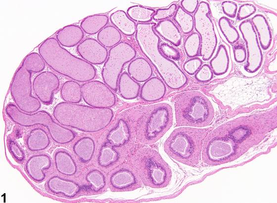
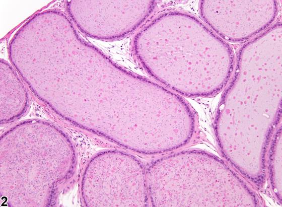
英文原文
展开 NTP 官方英文描述与建议
Comment:
Reduced sperm content is generally caused by reduced testicular spermatogenesis but can also be due to an obstructive lesion in the efferent ducts preventing sperm from a normal-appearing testis from reaching the epididymis. Sperm normally reach the cauda in 3-5 days and remain stored there for a week or more. Some toxicants can cause a decrease in transit time, thereby reducing normal sperm content (with no effect on spermatogenesis). Reduced testosterone levels will also result in reduced survival of sperm in the epididymis. A reduction in sperm is often accompanied by an increase in sloughed testicular germ cells and cell debris, due to associated spermatogenic disturbances in the testis. A reduction in sperm content is also often associated with a reduction in the size of the ductal lumen (ductal atrophy) due to the reduced volume of sperm and fluid. There may be a reduction (oligospermia) or a total absence (aspermia) of sperm; the degree of reduction can be represented using severity grades. Peripubertal rats (8-10 weeks) and mice (5-7 weeks) may have normal-appearing testes, normal amounts of sperm in the caput and corpus, but reduced amounts of sperm in the cauda because they have not built up full sperm reserves. This should not be considered abnormal in animals of this age.
Recommendation:
Reduced sperm content should be recorded and graded and should be discussed in the pathology narrative if the incidence and/or severity appears to be related to chemical administration. Bilateral involvement should be recorded when present. It is important to compare any potential changes with age-matched controls and to correlate with any available sperm count data. Any correlation with disturbances in testicular spermatogenesis should also be noted in the pathology narrative to aid interpretation. Correlation with any spermatogenic disturbances should also be noted in the pathology narrative to aid interpretation.
来源与引用
原始条目:https://ntp.niehs.nih.gov/atlas/nnl/reproductive-system-male/epididymis/Hypospermia
图谱总目录:NTP Nonneoplastic Lesion Atlas。 内容属美国联邦政府作品(公有领域),可引用;引用时请标注来源。
建议引用格式:NTP Nonneoplastic Lesion Atlas. Hypospermia (epididymis). LabSmart 中文版:
https://www.labsmart.cn/ntp/epididymis-hypospermia.html
中文图谱总页(可展开对照全部 595 条):https://www.labsmart.cn/ntp-atlas.html#epididymis-hypospermia