精液囊肿Spermatocele · 附睾 · 雄性生殖系统 · NTP ntp-0504
诊断要点:精液囊肿应记录为存在(present),并且如果其发生率似乎与化学物给药相关,则应在病理学叙述中加以讨论。
病变描述
精液囊肿(spermatocele)是附睾管的囊性扩张(直径达到正常管的3倍或以上),其内衬可为肥大的或正常的上皮。它们最常见于小鼠附睾的起始段或头区(Figure 1),内含数量不等的精子。该精液囊肿内可见局灶性(focal)矿化区(箭头,Figure 1)。精液囊肿通常作为背景性、偶然性病变出现,但也有报道其与某些化学物暴露相关。其与精子肉芽肿的鉴别在于无炎症反应。
诊断与分级建议(NTP 共识)
精液囊肿应记录为存在(present),并且如果其发生率似乎与化学物给药相关,则应在病理学叙述中加以讨论。如果双侧附睾均存在,诊断应限定为双侧性(双侧(bilateral))。不必对精液囊肿进行分级。
图谱
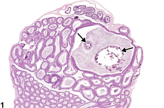
英文原文
展开 NTP 官方英文描述与建议
Comment:
Spermatoceles are cystic dilations of the epididymal duct (three or more times the diameter of the normal duct), which may be lined by hypertrophic or normal epithelium. They are most commonly seen in the initial segment or the caput region of the mouse epididymis (Figure 1) and are filled with varying amounts of sperm. Focal areas of mineralization are present within this spermatocele (arrows, Figure 1 ). Spermatoceles generally occur as background, incidental lesions but have been reported in association with exposure to some chemicals. They are distinguished from sperm granuloma by the absence of an inflammatory response.
Recommendation:
Spermatoceles should be recorded as present and discussed in the pathology narrative if the incidence appears to be related to chemical administration. If present in both epididymides, the diagnosis should be qualified as bilateral. It is not necessary to grade spermatoceles.
来源与引用
原始条目:https://ntp.niehs.nih.gov/atlas/nnl/reproductive-system-male/epididymis/Spermatocele
图谱总目录:NTP Nonneoplastic Lesion Atlas。 内容属美国联邦政府作品(公有领域),可引用;引用时请标注来源。
建议引用格式:NTP Nonneoplastic Lesion Atlas. Spermatocele (epididymis). LabSmart 中文版:
https://www.labsmart.cn/ntp/epididymis-spermatocele.html
中文图谱总页(可展开对照全部 595 条):https://www.labsmart.cn/ntp-atlas.html#epididymis-spermatocele