炎症Inflammation · 眼 · 特殊感觉系统 · NTP ntp-0330
诊断要点:眼部炎症只要存在就应诊断并分级。炎症应对每个眼部结构(例如角膜、前房、虹膜、睫状体、视网膜)分别诊断。
病变描述
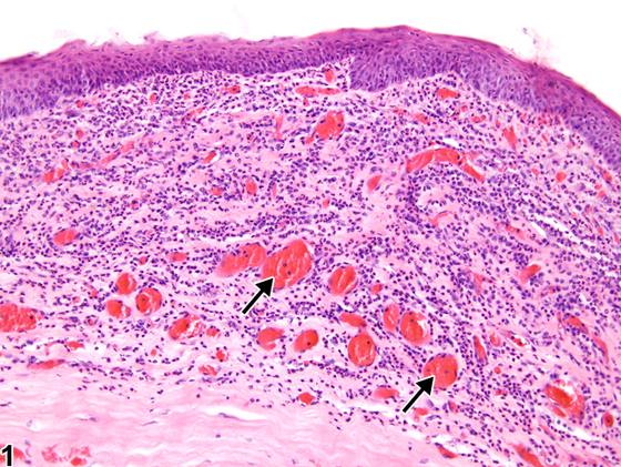
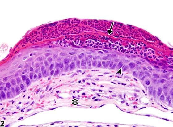
除角膜可能例外,眼部炎症是一种不常见的背景性病变。它可以发生在任何眼部结构中,并且常常同时出现于多个眼部结构。眼部炎症有多种原因,包括外伤(尤其是角膜)、化学刺激物和全身性毒素。角膜炎症可以是弥漫性的,也可以局限于特定区域,例如上皮或基质。除上述原因外,角膜炎症还可由泪液分泌不足、营养缺乏以及感染因子(如细菌、真菌和病毒)引起。由于角膜通常是无血管组织,基质新生血管形成是角膜炎症性病变的常见组成部分。上皮增生也是常见的表现(Figure 1、Figure 2)。在慢性病变中,可见基质纤维化。虽然完整的角膜对大多数细菌性病原体具有抵抗力,但上皮的任何破损(例如由擦伤、穿透性损伤或毒素诱导的坏死引起)都可能引入细菌,从而引发炎症反应。细菌还可继发性定植于原有的炎症区域(Figure 3、Figure 4)和/或溃疡区域。前房炎症可延伸至邻近的虹膜、睫状体和晶状体。前房中的炎症细胞可阻塞虹膜角膜滤角通道和/或邻近的小梁网,阻碍房水的正常引流,从而导致眼内压升高。临床上,前房内中性粒细胞积聚称为前房积脓(hypopyon)。前葡萄膜束(虹膜和睫状体)炎症可由多种原因引起,例如细菌和病毒等感染因子,以及免疫介导(过敏性)反应。虹膜和睫状体炎症(Figure 7、Figure 8, and Figure 9)常延伸至邻近的前房和后房、角膜和/或脉络膜。同样,脉络膜(后葡萄膜)炎症(Figure 10、Figure 11)常延伸至前葡萄膜束(睫状体和虹膜)和/或邻近的视网膜。玻璃体炎症(Figure 12、Figure 13)的特征是玻璃体内有明显的炎症细胞积聚。玻璃体炎症可由外伤(例如玻璃体内注射或其他穿透性损伤)引起,也可作为玻璃体内或全身给予毒素的原发效应而发生。它也可继发于其他眼部病变,如白内障和视网膜炎症。慢性炎症性浸润常伴有玻璃体纤维化。球后炎症(Figure 14、Figure 15)最常由球后出血性操作所致的外伤引起,通常为急性。它常伴有出血。在NTP研究中,炎症有五个标准类别:急性、化脓性、慢性、慢性活动性和肉芽肿性。在急性炎症中,主要浸润细胞是中性粒细胞,尽管也可能存在较少的巨噬细胞和淋巴细胞。还可能有水肿或充血的证据。中性粒细胞也是化脓性炎症中的主要浸润细胞类型,然而,在化脓性炎症中,中性粒细胞聚集,其中许多发生退变(化脓性渗出物)。渗出物中还可能存在来自驻留细胞群和浸润白细胞的细胞碎片、含纤维蛋白的蛋白性液体、较少的巨噬细胞、偶见的淋巴细胞或浆细胞,以及可能的感染因子。肉眼上,这些病变的特征是存在脓液。在渗出物周围的组织中,可能存在成纤维细胞、纤维结缔组织和混合性炎症细胞,具体取决于病变的慢性程度。淋巴细胞在慢性炎症中占主导地位。淋巴细胞在慢性活动性炎症中也占主导地位,但在慢性活动性炎症中,还有大量中性粒细胞。两种病变都可能含有巨噬细胞。肉芽肿性炎症是慢性炎症的另一种形式,但该诊断要求存在大量聚集的、大的、活化的巨噬细胞、上皮样巨噬细胞或多核巨细胞。炎症与细胞性浸润的鉴别在于存在其他变化,如水肿、出血、变性、坏死或其他组织损伤的证据。
诊断与分级建议(NTP 共识)
眼部炎症只要存在就应诊断并分级。炎症应对每个眼部结构(例如角膜、前房、虹膜、睫状体、视网膜)分别诊断。如果炎症位于球后间隙,应使用修饰词“retrobulbar”。诊断中应包含适当的类型修饰词(急性、慢性等)。两个或更多亚部位(例如睫状体和虹膜)不应在同一行条目诊断中使用。相关病变,如纤维化或新生血管形成,除非因严重程度而有必要,不应单独诊断,但应在病理学叙述中描述。
图谱
英文原文
展开 NTP 官方英文描述与建议
Comment:
With the possible exception of the cornea, ocular inflammation is an uncommon background lesion. It can occur in any of the ocular structures and often present in multiple ocular structures concurrently. Ocular inflammation has a number of causes, including trauma (especially the cornea), chemical irritants, and systemic toxins. Corneal inflammation can be diffuse or localized in particular zones, such as the epithelium or stroma. In addition to the causes listed above, corneal inflammation can also be caused by insufficient tear production, nutritional deficiencies, and infectious agents, such as bacteria, fungi, and viruses. Since the cornea is normally an avascular tissue, stromal neovascularization is a common component of corneal inflammatory lesions. Hyperplasia of the epithelium is also a frequent finding ( Figure 1 and Figure 2 ). In chronic lesions, stromal fibrosis may be seen. While the intact cornea is resistant to most bacterial pathogens, any breach in the epithelium (e.g., from abrasions, penetrating injury, or toxin-induced necrosis) can introduce bacteria, which incite an inflammatory response. Bacteria can also secondarily colonize preexisting areas of inflammation ( Figure 3 and Figure 4 ) and/or ulceration. Anterior chamber inflammation can extend into the adjacent iris, ciliary body, and lens. Inflammatory cells in the anterior chamber can occlude the iridocorneal filtration angle channels and/or the adjacent trabecular meshwork, impeding normal drainage of aqueous humor with resultant elevations in intraocular pressure. Clinically, intracameral accumulations of neutrophils are referred to as hypopyon. Anterior uveal tract (iris and ciliary body) inflammation can result from various causes, such as infectious agents such as bacteria and viruses, and immune-mediated (allergic) reactions. Iridial and ciliary body inflammation ( Figure 7 , Figure 8 , and Figure 9 ) often extends into the adjacent anterior and posterior chambers, cornea, and/or choroid. Likewise, inflammation of the choroid, or posterior uvea ( Figure 10 and Figure 11 ), often extends into the anterior uveal tract (ciliary body and iris) and/or into the adjacent retina. Vitreal inflammation ( Figure 12 and Figure 13 ) is characterized by prominent accumulations of inflammatory cells in the vitreous. Vitreal inflammation can result from trauma (e.g., from intravitreal injections or other penetrating injury) or as a primary effect of intravitreally or systemically administered toxins. It can also occur secondary to other ocular lesions, such as cataracts and retinal inflammation. Chronic inflammatory infiltrates are often accompanied by vitreal fibrosis. Retrobulbar inflammation ( Figure 14 and Figure 15 ) is most frequently caused by trauma due to retrobulbar bleeding procedures and is typically acute. It is often accompanied by hemorrhage. In NTP studies, there are five standard categories of inflammation: acute, suppurative, chronic, chronic-active, and granulomatous. In acute inflammation, the predominant infiltrating cell is the neutrophil, though fewer macrophages and lymphocytes may also be present. There may also be evidence of edema or hyperemia. The neutrophil is also the predominant infiltrating cell type in suppurative inflammation, however, in suppurative inflammation, the neutrophils are aggregated and many of them are degenerate (suppurative exudate). Cell debris, both from the resident cell populations and infiltrating leukocytes, proteinaceous fluid containing fibrin, fewer macrophages, occasional lymphocytes or plasma cells, and, possibly, an infectious agent may also be present in within the exudate. Grossly, these lesions would be characterized by the presence of pus. In the tissue surrounding the exudate, there may be fibroblasts, fibrous connective tissue, and mixed inflammatory cells, depending on the chronicity of the lesion. Lymphocytes predominate in chronic inflammation. Lymphocytes also predominate in chronic-active inflammation, but in chronic-active inflammation, there are also a significant number of neutrophils. Both lesions may contain macrophages. Granulomatous inflammation is another form of chronic inflammation, but this diagnosis requires the presence of a significant number of aggregated, large, activated macrophages, epithelioid macrophages, or multinucleated giant cells. Inflammation is differentiated from cellular infiltrates by the presence of other changes, such as edema, hemorrhage, degeneration, necrosis, or other evidence of tissue damage.
Recommendation:
Inflammation in the eye should be diagnosed and graded whenever present. Inflammation should be diagnosed separately for each ocular structure (e.g., cornea, anterior chamber, iris, ciliary body, retina). The modifier as "retrobulbar" should be used if the inflammation is in the retrobulbar space. An appropriate type modifier (acute, chronic, etc.) should be included in the diagnosis. Two or more subtopographies (e.g., ciliary body and iris) should not be used in the same line-entry diagnosis. Associated lesions, such as fibrosis or neovascularization should not be diagnosed separately, unless warranted by severity, but should be described in the pathology narrative.
来源与引用
原始条目:https://ntp.niehs.nih.gov/atlas/nnl/special-senses-system/eye/Inflammation
图谱总目录:NTP Nonneoplastic Lesion Atlas。 内容属美国联邦政府作品(公有领域),可引用;引用时请标注来源。
建议引用格式:NTP Nonneoplastic Lesion Atlas. Inflammation (eye). LabSmart 中文版:
https://www.labsmart.cn/ntp/eye-inflammation.html
中文图谱总页(可展开对照全部 595 条):https://www.labsmart.cn/ntp-atlas.html#eye-inflammation