晶状体白内障Lens Cataract · 眼 · 特殊感觉系统 · NTP ntp-0329
诊断要点:白内障应被诊断并赋予严重程度分级。诊断中应包含部位修饰词“lens”(即 Eye, Lens - Cataract)。
病变描述
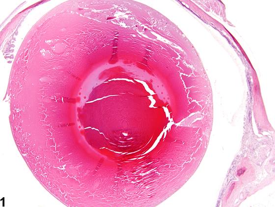
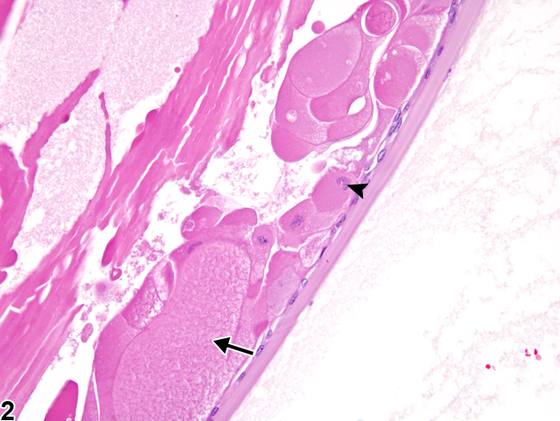
白内障(cataract)是晶状体混浊(囊性、囊下、前皮质或后皮质或核性),可由任何原因引起,包括遗传、创伤、代谢性疾病、营养失衡、环境因素、眼内压升高、衰老以及过度暴露于电离辐射或环境光。白内障也可与其他眼部疾病并发或继发于其他眼部疾病,如严重炎症、虹膜粘连和/或视网膜变性。病因不明、偶然发生的自发性白内障在老龄大鼠和小鼠中并不少见。白内障的特征是晶状体纤维具有各种异常特征,包括分离、肿胀、颗粒化、浓缩、断裂和正常有序结构的破坏,以及数个肿胀晶状体纤维中晶状体纤维核的异常滞留(Figure 1、Figure 2)。伴有异常滞留核的肿胀晶状体纤维有时被称为“膀胱”细胞或“气球”细胞。白内障还可表现出其他特征,如囊膜增厚、皱褶和/或破裂(Figure 3)或晶状体纤维或囊膜矿化(Figure 4)。白内障晶状体的前上皮细胞也可在损伤刺激下发生增殖和纤维化生(上皮-间充质转化)。白内障晶状体中还可能有前囊下纤维化(Figure 5)(注意混杂的深嗜酸性、碎裂的晶状体纤维物质),以及前上皮增生(Figure 6)。增殖的前上皮还可延伸至晶状体弓后方(晶状体上皮后移)(Figure 6)。
诊断与分级建议(NTP 共识)
白内障应被诊断并赋予严重程度分级。诊断中应包含部位修饰词“lens”(即 Eye, Lens - Cataract)。如果与治疗效应的特征描述相关,白内障的亚定位(囊下、皮质、核性等)应在病理学叙述中描述,但不应包含在诊断中。白内障的形态学特征(矿化、纤维化生、膀胱细胞等)不应单独诊断,但也应在叙述中描述。相关病变(如后粘连)应单独诊断。
图谱
英文原文
展开 NTP 官方英文描述与建议
Comment:
A cataract is a lens opacity (capsular, subcapsular, anterior or posterior cortical, or nuclear) resulting from any cause, including heredity, trauma, metabolic disease, nutritional imbalance, environmental factors, increased intraocular pressure, aging, and excessive exposure to ionizing radiation or ambient light. Cataracts can also be concurrent with or secondary to other ocular disease, such as severe inflammation, iridial synechiae, and/or retinal degeneration. Incidental, spontaneously occurring cataracts of uncertain etiology are not uncommon in aging rats and mice. Cataracts are characterized by lens fibers with various abnormal features, including separation, swelling, granularity, condensation, fragmentation, and disruption of the normally orderly configuration, as well as abnormal retention of lens fiber nuclei in several swollen lens fibers ( Figure 1 and Figure 2 ). Swollen lens fibers with abnormally retained nuclei are sometimes referred to as "bladder" or "balloon" cells. Cataracts can also exhibit additional features, such as capsular thickening, wrinkling, and/or rupture ( Figure 3 ) or lens fiber or capsular mineralization ( Figure 4 ). Anterior epithelial cells of cataractous lens can also undergo proliferation and fibrous metaplasia (epithelial-to-mesenchymal transition) in response to injury. There may also be anterior subcapsular fibrosis ( Figure 5 ) in a cataractous lens (note the intermixed dark eosinophilic, fragmented lens fiber material), as well as hyperplasia of the anterior epithelium of ( Figure 6 ). The proliferative anterior epithelium may also extend posterior to the lens bow (posterior migration of lens epithelium) ( Figure 6 ).
Recommendation:
Cataracts should be diagnosed and assigned a severity grade. The site modifier "lens" should be included in the diagnosis (i.e., Eye, Lens - Cataract). If pertinent to the characterization of a treatment effect, the subtopographical localization of cataracts (subcapsular, cortical, nuclear, etc.) should be described in the pathology narrative but should not be included in the diagnosis. Morphologic features of cataracts (mineralization, fibrous metaplasia, bladder cells, etc.) should not be diagnosed separately, but should also be described in the narrative. Associated lesions (e.g., posterior synechia) should be diagnosed separately.
来源与引用
原始条目:https://ntp.niehs.nih.gov/atlas/nnl/special-senses-system/eye/Lens-Cataract
图谱总目录:NTP Nonneoplastic Lesion Atlas。 内容属美国联邦政府作品(公有领域),可引用;引用时请标注来源。
建议引用格式:NTP Nonneoplastic Lesion Atlas. Lens Cataract (eye). LabSmart 中文版:
https://www.labsmart.cn/ntp/eye-lens-cataract.html
中文图谱总页(可展开对照全部 595 条):https://www.labsmart.cn/ntp-atlas.html#eye-lens-cataract