色素Pigment · 哈氏腺 · 特殊感觉系统 · NTP ntp-0319
诊断要点:由于卟啉色素是啮齿类动物眶外泪腺中的正常所见,只有当发生率或严重程度存在与受试物相关的差异时,才应对色素作出诊断并赋予严重程度分级。
病变描述
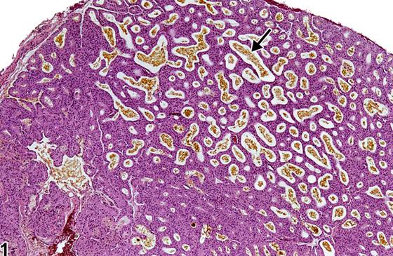
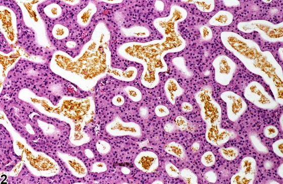
脂质是啮齿类动物眶外泪腺(Harderian gland)的主要分泌产物,但卟啉色素(porphyrin pigment)也会被合成并分泌。卟啉色素在腺泡腔内表现为金黄色至深棕色的无定形团块或层状凝结物(Figure 1、Figure 2)。卟啉分泌随年龄增长而增加,过度产生可导致“红泪”(chromodacyorrhea)。偶然发生的卟啉沉积通常在雌性大鼠和小鼠的眶外泪腺中更为显著。给予各种化学物质后,卟啉分泌增加也可能是与受试物相关的效应。
诊断与分级建议(NTP 共识)
由于卟啉色素是啮齿类动物眶外泪腺中的正常所见,只有当发生率或严重程度存在与受试物相关的差异时,才应对色素作出诊断并赋予严重程度分级。如果病理学家认为该色素由卟啉以外的物质(例如含铁血黄素(hemosiderin)或脂褐素(lipofuscin))构成,也应当作出诊断。即使使用一系列特殊染色,在组织学切片中确定色素的种类通常仍很困难。因此,推荐作出“色素”的诊断(而非诊断色素的类型,例如含铁血黄素或脂褐素),这是最恰当的做法。病理学描述应描述色素沉着的形态学特征。并非所有色素都必须作出诊断,因为有些色素在老龄动物中普遍存在,或与其他某种疾病过程相关,而不具有毒理学意义。病理学家应运用其判断力,决定继发性色素沉积是否显著到足以需要单独作出诊断。
图谱
英文原文
展开 NTP 官方英文描述与建议
Comment:
Lipids are the primary secretory products of the rodent Harderian gland, but porphyrin pigment is also synthesized and secreted. Porphyrin pigment occurs as golden to dark brown amorphous clumps or laminated concretions in the alveolar lumens ( Figure 1 and Figure 2 ). Porphyrin secretion increases with age, and excessive production results in "red tears" (chromodacyorrhea). Incidentally occurring porphyrin deposits are generally more prominent in the Harderian glands of female rats and mice. Increased porphyrin secretion can also be a treatment-related effect following administration of various chemical agents.
Recommendation:
Since porphyrin pigment is a normal finding in rodent Harderian glands, pigment should be diagnosed and assigned a severity grade only if there are treatment-related differences in incidence and/or severity. It should also be diagnosed if the pathologists feels the pigment is composed of something other than porphyrin (e.g., hemosiderin or lipofuscin). Definitive pigment identification is often difficult in histologic sections, even with a battery of special stains. Therefore, it is recommended that a diagnosis of pigment (as opposed to diagnosing the type of pigment, e.g., hemosiderin or lipofuscin) is most appropriate. The pathology narrative should describe the morphologic features of the pigmentation. Not all pigments have to be diagnosed, as some are ubiquitous in aging animals or related to some other disease process and not toxicologically meaningful. The pathologist should use his or her judgment in deciding whether or not secondary deposits of pigment are prominent enough to warrant a separate diagnosis.
来源与引用
原始条目:https://ntp.niehs.nih.gov/atlas/nnl/special-senses-system/harderian-gland/Pigment
图谱总目录:NTP Nonneoplastic Lesion Atlas。 内容属美国联邦政府作品(公有领域),可引用;引用时请标注来源。
建议引用格式:NTP Nonneoplastic Lesion Atlas. Pigment (harderian-gland). LabSmart 中文版:
https://www.labsmart.cn/ntp/harderian-gland-pigment.html
中文图谱总页(可展开对照全部 595 条):https://www.labsmart.cn/ntp-atlas.html#harderian-gland-pigment