矿化Mineralization · 肾脏 · 泌尿系统 · NTP ntp-0159
诊断要点:矿化应当诊断并分级。如果小的局灶性(focal)矿化沉积物被认定为自发性背景性病变,则无需诊断,病理学家应运用其判断来决定这些沉积物是否足够显著而值得诊断。
病变描述
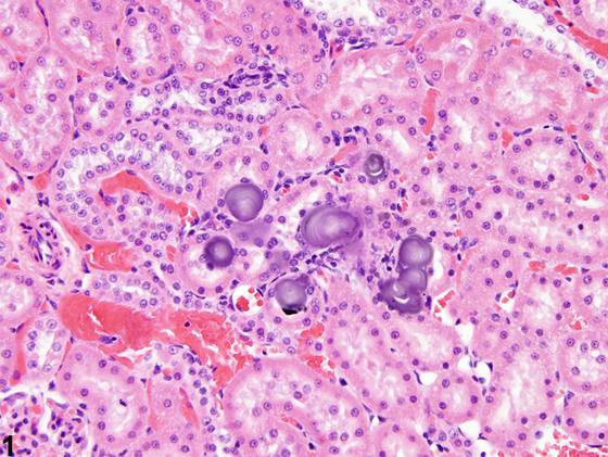
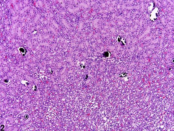
矿化更常与大鼠和小鼠肾皮质、髓质或肾乳头中嗜碱性沉积物的自发性、微小背景性病变相关(Figure 1)。一般而言,这些沉积物无病理学意义。然而,矿化也可见于变性和坏死的继发结果。矿化可由化学物、激素或饮食诱导。矿化沉积物通常累及肾小管上皮,但也可能累及间质。茜素红S(Alizarin red S)或von Kossa特殊染色用于在组织学切片中显示矿物质。矿化沉积物尤其见于雌性大鼠,位于外髓外带与外髓内带交界处附近的直部(pars recta)和Henle细环(Figure 2)。肾乳头矿化的线性沉积是诱导α2u-球蛋白肾病(α2u-globulin nephropathy)的慢性化学物给药的特征性表现(Figure 3)。外皮质有时可见人为假象性矿化。这种矿化的原因通常无法确定。
诊断与分级建议(NTP 共识)
矿化应当诊断并分级。如果小的局灶性(focal)矿化沉积物被认定为自发性背景性病变,则无需诊断,病理学家应运用其判断来决定这些沉积物是否足够显著而值得诊断。当进行诊断时,矿化的模式(例如,线性肾乳头矿化、局灶性髓质矿化)应在病理学描述中予以描述。相关链接:肾脏-结石 肾脏-结晶
图谱
英文原文
展开 NTP 官方英文描述与建议
Comment:
Mineralization is more commonly associated with spontaneous and minute background findings of basophilic deposits in the renal cortex, medulla,or papilla of rats and mice ( Figure 1 ). In general, these depositshave no pathologic significance. However, mineralization may also be seen as a consequence to degeneration and necrosis. Mineralization may be induced by chemicals, hormones, or diet. Mineralized deposits usually involve tubule epithelia but may also involve the interstitium. Alizarin red S or von Kossa’s special stains are used to demonstrate mineral in histologic sections. Mineralized deposits seen particularly in female rats are noted in the pars recta and thin loops of Henle near the junction of the outer stripe of the outer medulla and the inner stripe of the outer medulla ( Figure 2 ). Linear deposits of papillary mineralization are characteristic of chronic chemical administration that induces α2u-globulin nephropathy ( Figure 3 ). Artifactual mineralization is sometimes encountered in the outer cortex. The cause of this mineralization often cannot be identified.
Recommendation:
Mineralization should be diagnosed and graded. If small deposits of focal mineralization are recognized as a spontaneous background finding, they need not be diagnosed and the pathologist should use his or her judgment in deciding whether or not they are prominent enough to warrant diagnosis. When diagnosed, the pattern of the mineralization (e.g., linear papillary mineralization, focal medullary mineralization) should be described in the pathology narrative. Related Links: Kidney - Calculus Kidney - Crystals
来源与引用
原始条目:https://ntp.niehs.nih.gov/atlas/nnl/urinary-system/kidney/Mineralization
图谱总目录:NTP Nonneoplastic Lesion Atlas。 内容属美国联邦政府作品(公有领域),可引用;引用时请标注来源。
建议引用格式:NTP Nonneoplastic Lesion Atlas. Mineralization (kidney). LabSmart 中文版:
https://www.labsmart.cn/ntp/kidney-mineralization.html
中文图谱总页(可展开对照全部 595 条):https://www.labsmart.cn/ntp-atlas.html#kidney-mineralization