肾母细胞瘤病Nephroblastematosis · 肾脏 · 泌尿系统 · NTP ntp-0180
诊断要点:肾母细胞瘤病一旦存在即应诊断,并给出严重程度分级。
病变描述
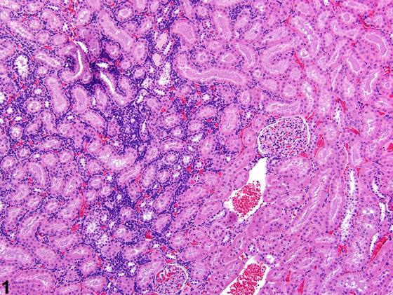
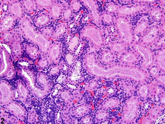
肾母细胞瘤病(nephroblastematosis)可为自发性病变,是肾母细胞瘤(nephroblastoma)的假定前驱病变。低倍镜下,肾母细胞瘤病表现为细胞密集的嗜碱性区域(Figure 1、Figure 2)。高倍镜下,可见形态一致的细胞浸润于肾小管之间,其特征为细胞边界和胞质不清,细胞核呈圆形至卵圆形,染色质呈点状(Figure 3)。肾母细胞瘤病与肾母细胞瘤之间的区分在一定程度上是人为的,取决于是否分化形成任何器官样(小管、肾小球样)结构——该结构见于肾母细胞瘤病——以及病变的整体大小。与其他病变(如细胞浸润、炎症或淋巴瘤)的鉴别通常取决于是否存在细胞类型更为异质性的细胞群(常呈局灶性(focal)至多灶性(multifocal)血管周围分布)和/或病变内血管中是否存在相关细胞(Figure 4)。
诊断与分级建议(NTP 共识)
肾母细胞瘤病一旦存在即应诊断,并给出严重程度分级。
图谱
英文原文
展开 NTP 官方英文描述与建议
Comment:
Nephroblastematosis can be a spontaneous lesion and is the presumptive precursor lesion of nephroblastoma. On low power, nephroblastematosis appears as an area of densely basophilic cellularity ( Figure 1 and Figure 2 ). On higher magnification, uniform cells are seen infiltrating between renal tubules and are characterized by having indistinct cell margins and cytoplasm and round to oval nuclei with stippled chromatin ( Figure 3 ). The distinction between nephroblastematosis and nephroblastoma is somewhat arbitrary and depends on differentiation into any organoid (tubule, glomeruloid) structures, which is seen in nephroblastematosis, and the overall size of the lesion. Differentiation from other lesions such as cellular infiltrates, inflammation, or lymphoma usually depends on the presence of a more heterogeneous population of cell types, which often have a focal to multifocal perivascular distribution, and/or the presence of associated cells in blood vessels within the lesion ( Figure 4 ).
Recommendation:
Nephroblastematosis should be diagnosed whenever present and given a severity grade.
来源与引用
原始条目:https://ntp.niehs.nih.gov/atlas/nnl/urinary-system/kidney/Nephroblastematosis
图谱总目录:NTP Nonneoplastic Lesion Atlas。 内容属美国联邦政府作品(公有领域),可引用;引用时请标注来源。
建议引用格式:NTP Nonneoplastic Lesion Atlas. Nephroblastematosis (kidney). LabSmart 中文版:
https://www.labsmart.cn/ntp/kidney-nephroblastematosis.html
中文图谱总页(可展开对照全部 595 条):https://www.labsmart.cn/ntp-atlas.html#kidney-nephroblastematosis