肾小管坏死Renal Tubule Necrosis · 肾脏 · 泌尿系统 · NTP ntp-0172
诊断要点:坏死应当被诊断并给予严重程度分级。与肿瘤存在相关的坏死不应被诊断,而应在病理学叙述中描述。
病变描述
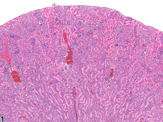
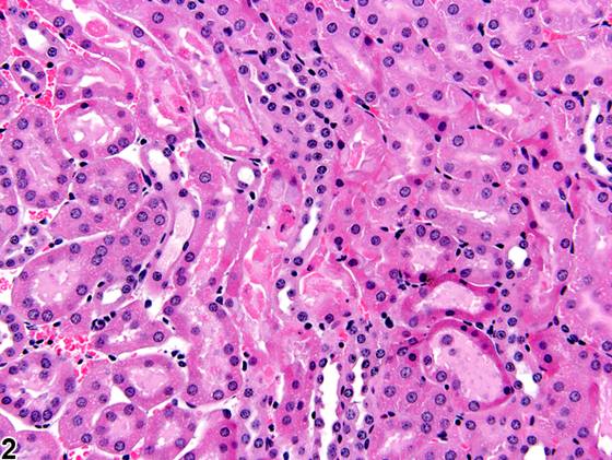
坏死的诊断涵盖单个细胞死亡、局灶性(focal)或多灶性(multifocal)的肾小管坏死区域,直至较大融合区域的肾实质坏死(Figure 1)。坏死可见于肾脏的任何部位。典型表现为细胞肿胀、细胞碎裂、胞质染色性改变和核碎裂(Figure 2)。坏死的肾小管细胞迅速脱落进入肾小管腔。肾小管内的颗粒管型是既往坏死的指示性表现。急性肾小管坏死时很少观察到炎症。
诊断与分级建议(NTP 共识)
坏死应当被诊断并给予严重程度分级。与肿瘤存在相关的坏死不应被诊断,而应在病理学叙述中描述。继发性改变,如再生和炎症,不应单独诊断,除非严重程度足以支持,但应在叙述中描述。肾小管坏死必须与人为假象相鉴别。相关链接:肾脏 - 肾小管 - 变性;肾脏 - 肾小管 - 再生
图谱
英文原文
展开 NTP 官方英文描述与建议
Comment:
The diagnosis of necrosis encompasses death of individual cells, to focal or multifocal areas of tubule necrosis, to larger confluent areas of renal parenchyma necrosis ( Figure 1 ). Necrosis may be seen anywhere in the kidney. Typically, it is characterized by cell swelling, cell fragmentation, cytoplasmic tinctorial changes, and karyorrhexis ( Figure 2 ). Necrotic renal tubule cells quickly slough off into the tubule lumens. Granular casts within renal tubules are indicators of previous necrosis. Inflammation is seldom observed in association with acute tubule necrosis.
Recommendation:
Necrosis should be diagnosed and given a severity grade. Necrosis associated with the presence of a tumor should not be diagnosed but should be described in the pathology narrative. Secondary changes, such as regeneration and inflammation, should not be diagnosed separately, unless warranted by severity, but should be described in the narrative. Renal tubular necrosis must be differentiated from artifact. Related Links: Kidney - Renal Tubule - Degeneration Kidney - Renal Tubule - Regeneration
来源与引用
原始条目:https://ntp.niehs.nih.gov/atlas/nnl/urinary-system/kidney/RenalTubule-Necrosis
图谱总目录:NTP Nonneoplastic Lesion Atlas。 内容属美国联邦政府作品(公有领域),可引用;引用时请标注来源。
建议引用格式:NTP Nonneoplastic Lesion Atlas. Renal Tubule Necrosis (kidney). LabSmart 中文版:
https://www.labsmart.cn/ntp/kidney-renal-tubule-necrosis.html
中文图谱总页(可展开对照全部 595 条):https://www.labsmart.cn/ntp-atlas.html#kidney-renal-tubule-necrosis